
Page 207 - 2020_02-Haematologica-web
P. 207
High STAT5A activity promotes CD8+ T-cell neoplasia
or 30 genes (10.5%) were specifically upregulated in cS5Ahi vs. wt or cS5Ahi vs. cS5Alo cells, respectively. Most downregulated genes (253 genes, 65.2%) were seen in cS5Ahi compared to wt mice, whereas only 11 genes (2.8%) showed lowered expression in cS5Ahi compared to cS5Alo animals (Figure 5A, Online Supplementary Figure S5A). We confirmed differential expression of well- described STAT5-target genes Pim1, Bcl2, Bcl6 and Cish by qRT-PCR (Online Supplementary Figure S5B). Gene set enrichment analysis on genes significantly up- or down- regulated in wt vs. cS5Ahi or cS5Alo vs. cS5Ahi mice con- firmed the IL-2-STAT5 signaling axis and revealed enrich- ment of E2F and Myc targets and G2M checkpoint genes as well as a lowered interferon (IFN) response in STAT5 hyperactive mice (Figure 5B, Online Supplementary Figure S5C). This matches the described STAT5-IFN axis in transformation.45 Stat5a and Stat5b share very similar roles in T cells.46 However, sequencing efforts attribute an important role to the activating STAT5BN642H variant.28,32 To compare the phenotypically largely overlapping, though much more aggressive, disease of hSTAT5BN642H and cS5Ahi mice, we contrasted gene expression patterns of wt, cS5Alo, cS5Ahi, hSTAT5B and hSTAT5BN642H CD8+ T cells (Figure 5C, Online Supplementary Figure S5D, RNA- sequencing of hSTAT5B and hSTAT5BN642H as published32). The hSTAT5B and cS5Alo expression profiles cluster with that of wt T cells (Online Supplementary Figure S5D), whereby hyperactive STAT5A and STAT5B signaling share 373 (28.8%) commonly deregulated genes (Figure
5D). Importantly, both CD8+ T-cell neoplasia models are enriched for genes reported to be altered in PTCL, NOS with cytotoxic T-cell features6,7 (Figure 5E and Online Supplementary Table S5), the closest match being to dereg- ulated cS5Ahi genes in the tested T-cell lymphoma gene sets (Online Supplementary Figure S5E). Enrichr pathway analysis47 of the shared cS5Ahi and hSTAT5BN642H gene expression signature revealed an upregulation of cytokine-cytokine receptor interaction and JAK/STAT sig- naling. The exclusive hSTAT5BN642H genes can be attributed to enhanced cell cycle and division, which may explain its sensitivity to Aurora Kinase inhibition32 (Online Supplementary Figure S5F, Online Supplementary Table S6).
We conclude that the immunophenotype, pathology and gene signatures of the cS5Ahi-induced PTCL-like dis- ease overlap with those of human PTCL, NOS with cyto- toxic T-cell features, which is associated with a particular- ly poor prognosis. hSTAT5BN642H correlates similarly but is more aggressive. This implies that a significant threshold of STAT5 activity is not only required to induce, but is also sufficient to promote PTCL development.
Elevated STAT5 activation in human mature T-cell lymphomas
Subsequently, we investigated STAT5A or STAT5B expression and cellular localization in PTCL entities by specific immunohistochemical STAT5A or STAT5B stain- ing. Quantification of nuclear STAT5 staining intensity revealed that PTCL, NOS cases had higher expression
AD
B
C
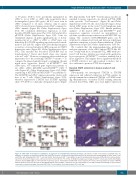
Figure 4. cS5Ahi CD8+ T-cell transfer recapitulates an aggressive T-cell lymphoma/leukemia phenotype. (A) Scheme of CD8+ T-cell transfer. LN: lymph node; wt: wildtype; i.v.: intravenous. (B) Representative flow cytometry dot plots of recipient’s peripheral blood 12 weeks after injection showing gating on CD3e+ CD8+ T cells and further Ly5.1/2 gating (left). CD8+ T cells and %Ly5.2+ donor-derived CD8+ T cells (right) in recipient’s peripheral blood measured after injection at 4-week inter- vals for 16 weeks (n=6/organ source, untreated control n=2, wt LN n=1, P<0.0001). (C) Endpoint analysis: percentage of CD8+ T cells in spleen (left), LN (middle) and bone marrow (BM) (right) of control (wt-CD8+ recipients), spleen- and LN-derived cS5Ahi-CD8+ T-cell recipients (P<0.0001). Two-way analysis of variance (ANOVA) with the Bonferroni post test (B) and one-way ANOVA with the Tukey multiple comparison test. (D) Hematoxylin & eosin (HE) and anti-CD3 staining of representative consecutive sections from lungs of wt and cS5Ahi-CD8+ T-cell recipients. Scale bars indicate 50 or 100 μm.
haematologica | 2020; 105(2)
441
