
Page 116 - 2020_02-Haematologica-web
P. 116
G. Goyal et al.
macroglobulinemia). Immune-related RDD was diag- nosed in five patients, with one case each of rheumatoid arthritis, multiple sclerosis, Sjögren's syndrome, systemic lupus erythematosus, and warm autoimmune hemolytic anemia. Serologic evaluation was not indicated in the remaining patients due to a lack of clinical features of con- comitant autoimmune disorders. Three patients had high IgG4 level expression in lesional lymphoplasmacytic cells on immunohistochemistry, but only one had elevated serum IgG4 levels. None of these patients had other fea- tures consistent with IgG4-related disease.
Organ involvement
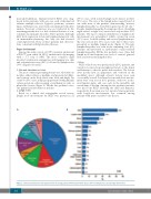
Among the entire cohort, 24 (38%) patients underwent a PET-CT scan, while 16 (25%) underwent body imaging with a CT scan or MRI. The most common organ involved on physical examination and imaging was skin and subcutaneous tissue (52%), followed by lymph nodes (33%) (Figures 1B and 2).
1. Skin and subcutaneous tissue
The most common presenting feature was subcutaneous nodules, either solitary or multiple, and presented at differ- ent locations on the body (chest, arm, back, and thigh). Six of the 33 (18%) cases in this group presented with primarily cutaneous lesions, either a purple or erythematous rash, or plaque-like lesions (Figure 3). Of the five pediatric cases, one patient had subcutaneous nodules.
2. Lymph nodes
Based on a clinical and radiographic record review, lymph node involvement by RDD was present in 21
AB
(33%) cases, with isolated lymph node disease in three (5%) cases. The size of the lymph nodes ranged from 1-2 cm, with none of the patients demonstrating “massive lymphadenopathy” as described in prior reports (≥7 cm).2,3 Despite lymphadenopathy, B-symptoms (fever, drenching night sweats, weight loss) were noted only in three (5%) patients. The most common distribution of lymph node involvement was generalized, which occurred in seven (11%) cases. Isolated axillary and cervical lymphadenopa- thy was seen in five (8%) cases each. All of these cases presented as multiple lymph nodes (Figure 4). Thoracic lymphadenopathy was seen in the remaining four (6%) patients, and presented as mediastinal or para-tracheal lymphadenopathy. Of the five pediatric cases, three had lymph node involvement (one each of cervical, general- ized, and retrocrural lymph nodes).
3. Bone
RDD of the bones was present in 16 (25%) patients, and varied in location from metaphyseal heads of the femur and humerus to the ribs, pelvis and vertebrae. The lesions were mostly lytic in appearance and centered in the medullary space, although sclerotic lesions were seen occasionally as well. Soft tissue lesions with bone involve- ment were seen in four (6%) patients, with two in tho- racic/lumbar spine, and one each in the mandible and acetabulum of the hip. Among the five pediatric patients, two had bone RDD involving the skull and humerus, respectively. Bone pain was not reported among patients with long-bone involvement, but common among patients with spine or pelvic bone disease.
Figure 1. Clinical manifestations and organ involvement among patients with Rosai-Dorfman disease A) Presenting features and B) Organ involvement
350
haematologica | 2020; 105(2)
