
Page 115 - 2020_02-Haematologica-web
P. 115
(LCH) has been enhanced by the discovery of recurrent BRAF and related mitogen activated protein kinase – extracellular signal-regulated kinase (MAP-ERK) pathway mutations.4-6 The identification of these specific muta- tions in both LCH and ECD further supported their con- sideration as neoplastic disorders rather than reactive inflammatory conditions. Recently, mutually exclusive KRAS and MAP2K1 mutations were identified in one- third of RDD patients, pointing toward a neoplastic process in this disease as well.7 Due to the rarity of RDD, the clinical spectrum and treatment outcomes are not well defined. Hence, we undertook this study to evaluate our institutional experience with RDD patients in a more contemporary setting.
Methods
The medical records of patients with RDD evaluated at a terti- ary referral center from January 1, 1994 to December 15, 2017 were identified and reviewed after approval from the Institutional Review Board. Definitive histopathological diagnosis by tissue biopsy review was necessary for inclusion in the study. All biop- sies identified at our institution (n=28) were re-reviewed by two pathologists with expertise in histiocytic disorders (K.L.R. and A.R.).8-10 Data abstracted from the medical records included: demographic characteristics, symptoms at disease presentation, histopathological features, treatment modalities utilized, and out- comes. In addition, radiologic and genomic findings were captured where available. Next generation sequencing (NGS) using an oncogene panel (FoundationOne® or Tempus®) was performed on five RDD tissue samples and one blood sample (Guardant360®).11- 13 All patients in the study were followed up by a medical record review until death or November 10, 2018, whichever was earlier. For patients that were lost to the follow up, additional information was acquired via a telephone interview and survey forms. To min- imize errors and bias, the medical records were independently reviewed by two investigators (GG and AR).
The majority of the patients did not undergo positron emis- sion tomography – computed tomography (PET-CT) scans for baseline evaluation or treatment-response assessment. Hence, we utilized data from the reports of imaging studies available — radiographs, CT scans, and magnetic resonance imaging (MRI) scans. The imaging studies selected to be included in the manu- script were reviewed independently by a radiologist with expertise in histiocytic disorders (JRY).14 RDD patients were classified into subgroups based on the location, as well as asso- ciated conditions.15 The sites of disease were based on histopathologic or radiographic findings and include those found at the follow-up as well. Based on the location, RDD involving the lymph nodes alone was classified as “classical” and others as “extranodal”. Based on consensus definitions of con- comitant disorders, RDD was classified as “neoplasia-associat- ed” RDD, “immune-related” RDD, and “IgG4-related” RDD.8
As there is no United States Food and Drug Administration (US-FDA) approved treatment for RDD, the patients were treat- ed with various therapeutic agents/modalities. We assessed treatment response by reviewing the clinical documentation. The response criteria were defined clinically and radiologically as we have previously described in ECD.16 Because RDD is a relapsing-remitting disease, we assessed the overall response rate (ORR), which incorporated complete as well as partial remissions (complete or partial resolution of symptoms or imag- ing finding suspected due to RDD). Descriptive statistics were used to summarize the data.
Results
Patient characteristics and presenting features
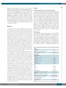
We included 64 RDD patients in the study. Of these, 8% had classical (nodal only) and 92% had extra-nodal RDD (67% extra-nodal only) (Table 1). Overall, 47 (73%) had multi-site disease and 17 (27%) had solitary or single-site disease. The median age at diagnosis was 50 years (range, 2-79). Five patients were less than 18 years of age (age 2, 2, 11, 14, and 15 years, respectively). In the entire cohort, there was a slight female preponderance (female: male 1.5:1). The median duration from symptom onset to diag- nosis was seven months (range, 0-128; mean 18 months). The most common presenting symptom was painful or painless subcutaneous masses (40%; Figure 1A). Symptoms due to lymphadenopathy were reported only in 11% of patients (Figure 1A). C-reactive protein level at diagnosis was available in 21 (33%) of patients, with a median value of 12 mg/L (range <0.3 to 198 mg/L; normal value <8 mg/L).
RDD subtypes
Overall, eight patients had neoplasia-associated RDD. Of these, three patients had RDD (two testicular, one vocal cord) in conjunction with ECD of other organ sys- tems, characterized as mixed or overlap histiocytosis. Three patients had RDD subsequent to the diagnosis of a hematologic malignancy (peripheral T-cell lymphoma not otherwise specified, marginal zone B-cell lymphoma, myelodysplastic syndrome with excess blasts-1), while two patients developed a hematologic malignancy after RDD diagnosis (mantle cell lymphoma, Waldenström's
Table 1. Clinical and baseline features of patients with Rosai-Dorfman disease.
Clinical features of Rosai-Dorfman disease
Total patients
RDD/ECD overlap (mixed histiocytosis) Median age at diagnosis (years) Female: Male
Classification
Familial
Classical (node-only) Extranodal Neoplasia-associated IgG4-related Immune-related
Race
White
Black
Asian Other/unknown
Median time from symptom to diagnosis
(months)
Median number of biopsies for diagnosis
Median duration of follow-up (months)
Median overall survival since onset of symptoms (months)
Lost to follow up
Deaths
64
3 (5%)
50 years (range, 2-79) 1.5:1
0
5 (8%) 59 (92%) 8 (13%) 3 (5%) 5 (8%)
40 (63%)
9 (14%)
3 (5%)
12 (18%)
7 (range, 0-128)
2 (range 1-6) 31 (range 0-249) 140 (range 8-684)
15 (23%)
4
haematologica | 2020; 105(2)
349
