
Page 118 - 2020_02-Haematologica-web
P. 118
G. Goyal et al.
4. Head and neck (including orbit)
Head and neck RDD lesions were noted in seven (11%) patients. Orbital involvement occurred in three (5%) cases, one of which was a pediatric patient. One of these also had ciliary body and scleral involvement. Other RDD sites in the head and neck region included the trachea (n=2), nose (n=1), and vocal cord (n=1).
5. Glandular tissue
RDD involving the glands was seen in nine cases, most common being breast tissue (n=5), with abnormalities on mammogram or MRI (Figure 5). Two patients each had involvement of lacrimal and parotid salivary gland with- out any evidence of dry eyes or mouth.
6. Kidneys, adrenals, abdomen and retroperitoneum
RDD of the kidneys was seen in six patients, most com- monly as solitary parenchymal mass or nodule, and less commonly as perinephric coating, without the classic “hairy kidney” appearance as seen with ECD.17 None of these patients had renal failure from RDD of the kidneys. Two of these patients had adrenal nodules. Other abdom- inal sites included mesentery and peritoneum in one patient each.
7. Nervous system
Central nervous system (CNS) involvement manifested as dural- or parenchymal-based lesions in four cases. Parenchymal lesions were observed on MRI imaging in
AD
three patients, manifesting as frontal or temporal solitary masses. One of these patients had pachymeningeal dis- ease along with cerebral subcortical white matter infiltra- tive lesions. Additionally, one patient had optic nerve involvement causing visual disturbance.
8. Cardiovascular and respiratory system
Cardiovascular involvement was uncommon, noted as a right atrial mass encasing the coronary artery in one patient and aortic infiltration in two patients (Figure 5). Pulmonary RDD was seen in four patients and presented as a parenchymal nodule, interstitial pneumonitis, or soli- tary pleura-based lesion.
9. Bone marrow, liver, and spleen
Biopsy proven bone marrow involvement was seen in one patient while three others had an increased bone mar- row signal on PET-CT. Liver involvement occurred in three cases and two had spleen lesions (Figure 5).
10. Other sites
RDD involving the testes was noted in three cases, two of which had ECD of other tissue sites (Figure 5). RDD of the maxillary and ethmoid sinuses was noted on CT scan of the head in four cases, with sinus-related symptoms in three patients. Two patients had paravertebral soft tissue nodules, one of whom presented with compression of the spinal cord from mass effect. Colon- and rectal-based polypoid lesions were found in two patients.
B
C
E
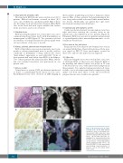
Figure 4. Rosai-Dorfman disease RDD) in lymph node. (A) The RDD infiltrate expands the sinuses of the lymph node. Characteristic RDD histiocytes show abundant cytoplasm with emperipolesis (inset). (B) The RDD histiocytes are highlighted by immunohistochemistry for S100, and (C) CD163. (D) Enhanced coronal thoracic CT depicting bilateral axillary lymphadenopathy (circle). (E) Fused FDG PET/CT of the same patient demonstrating hypermetabolism of bilateral cervical and axillary lymph nodes (circle).
352
haematologica | 2020; 105(2)
