
Page 99 - 2019_12-Haematologica-web
P. 99
Clonal hematopoiesis and AML risk
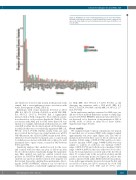
Figure 1. Distribution of exonic clonal mutations by gene in cases and controls.
Each bar represents the fraction of exonic mutations detected in each gene rel- ative to the total number of exonic mutations detected in cases (n=215) or con- trols (n=285).
ant, which we observed only in men in the present study sample, had a non-significant positive association with future development of AML (Table 2).
for AML (OR: 14.0, 95%CI: 1.7-113.8; P=0.01), as did detecting any mutation with a VAF ≥0.01 (OR: 5.1, 95%CI: 1.6-15.9; P=0.005) or ≥0.02 (OR: 5.3, 95%CI: 1.7- 16.4; P=0.004).
In the exploratory analyses restricted to AML cases, we did not observe marked differences in time to AML diag- nosis by DNMT3A R882H/C mutation status (detected vs. not detected) or by detection of any mutation at VAF of ≥0.005, ≥0.01 or ≥0.02 at either blood draw (Online Supplementary Figure S5A-D).
Clonal stability
We examined clonal evolution of mutations over time in 11 matched sets of women (NHS) with samples banked approximately ten years apart (Figure 2A). The VAF of mutations detected in these cases at blood collection one (median: 0.0021; range: 0.0003-0.0782) and blood collec- tion two (median: 0.0037; range: 0.0006-0.2992) was very similar to controls at collection one (median: 0.0017; range: 0.0003-0.0731) and collection two (median: 0.0023; range: 0.0003-0.2689). In the cases with two blood collec- tions, 31 clonal mutations occurred only at the first blood draw, 37 occurred only at the second blood draw, and 22 occurred at both time points (see Figure 2A; yellow data points connected with a line). Of the latter 22 clonal muta- tions, in the approximately ten years between the first and second blood draw, five (23%) increased by >0.01 VAF, none decreased by >0.01 VAF, and 17 (77%) were
Individuals with clonal mutations detected at ≥0.01 (OR: 5.4, 95%CI: 1.8-16.6; P=0.003) or ≥0.02 VAF (OR: 5.6, 95%CI: 1.8-17.2; P=0.003) had a significantly increased risk of AML compared to those without a muta- tion detected at or above those thresholds (Table 2). The association with AML risk for VAF lower than 0.01 was unclear; for example, individuals with mutations at a VAF of ≥0.005 at either blood collection had a 2.5-fold increase in AML risk that was not statistically significant (OR: 2.5, 95%CI: 1.0-6.3; P=0.05). Further, nearly every case and most controls had at least one clonal mutation at ≥0.001 VAF. Of interest, the ASXL1 E1183K variant noted above, which we observed in five women (4 cases, 1 control) and for which an association with AML risk could not be well quantified due to sparse counts, occurred at VAF between 0.001 and 0.002.
Sensitivity analyses that omitted records for the cases and controls with less than one year of follow up after blood collection did not materially change the main find- ings. One omitted case and two omitted controls were positive for DNMT3A R882H/C, whereas the remaining omitted case and four omitted controls were negative for that variant. Even after omitting these participants, detect- ing the DNMT3A R882H/C variant at one or both blood collections remained a statistically significant risk factor
haematologica | 2019; 104(12)
2413
