
Page 101 - 2019_12-Haematologica-web
P. 101
Clonal hematopoiesis and AML risk
In exploratory case-only analyses (see Online Supplementary Methods), the VAF for the most abundant clone observed at the first blood draw (i.e. the largest VAF observed at collection one) did not correlate to the time to diagnosis of AML (partial Spearman r = -0.11, P=0.55, adjusted for age and sex) (Figure 3A). In the NHS cases with a second collection blood sample, the maximum VAF at the second time point and time to AML diagnosis was not correlated (partial Spearman r=0.34, P=0.33, adjusted for age) (Figure 3B). The largest change in VAF between collections with time to AML diagnosis was also not cor- related (partial Spearman r=0.30, P=0.39, adjusted for age) (Figure 3C).
Discussion
In this study, we investigated associations of clonal hematopoiesis with long-term risk of AML, leveraging ECS-determined clonal variants and up to 22 years of fol- low up after blood draw in 34 matched case-control sets from the NHS and HPFS. Surprisingly, we found no clear differences in clonal mutation abundance, location or VAF between cases and controls. As expected, DNMT3A and TET2 were the genes with the most frequently detected clonal mutations in both cases and controls,2,6,14 and over- all, cases and controls showed abundant mutation across the rest of the coding sequence. Few individual variants
occurred frequently enough for separate analysis of AML risk, but among those occurring in at least four partici- pants, DNMT3A R882H/C had a strong association with AML risk. We also observed statistically significant associ- ations with AML risk for individuals with any variant with a VAF ≥0.01. Contrary to expectation, in the 11 matched sets with two banked blood samples, we did not observe a signature of clonal evolution over time that dis- tinguished cases from controls or predicted latency to AML diagnosis in the cases.
Two recent studies reported findings for clonal hematopoiesis and future risk of AML.4,5 Briefly, both studies observed an increased risk of AML for increasing numbers of clonal mutations, higher VAF and detection or number of mutations in known driver genes. Of note, Abelson et al.5 observed an increased AML risk for individ- uals with clones of VAF ≥0.005 detected by ECS, and Desai et al.4 reported an increased risk of AML for women with clones of VAF ≥0.01 detected by targeted deep sequencing. We detected an increase in AML risk for per- sons with clonal mutations at ≥0.01 VAF and those with DNMT3A R882 mutations, and our observed effect esti- mates had a similar magnitude and precision as those reported by the previous studies. Other prior studies reported that these mutations raise AML risk by 0.5- 1%/year.2,3 For mutations detected at VAF <0.01, our find- ings were less clear due to limited statistical precision. Additionally, in the subset of women with repeat blood
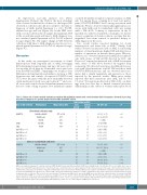
Table 2. Future risk of acute myeloid leukemia associated with individual variants and selected variant allele frequencies detected in pre-diag- nosis blood samplesa in a pooled sample from the NHS and HPFS cohorts.
Gene or VAF criterionb Polymorphism
Major/minor allele
Total testing positivea Cases Controls (N=34)c (N=69)c
7 3
OR (95% CI)d P
6.3 (1.3, 30.7) 0.02 12.0 (1.4, 99.7) 0.02
DNMT3A
R882H or R882C R882H only
C/T or G/A C/T
Any VAF ≥0. 001 Any VAF ≥0.005 Any VAF ≥0.01 Any VAF ≥0.02
DNMT3A
First blood collection only
6 1
W860R A/G 0 4 0.0(NC)
ASXL1
JAK2 V617F G/T 4e 1e
2.0x10⁷(NC) 8.0 (0.9, 71.6) 0.06 2.3x10⁷ (NC) 2.4 (1.0, 6.1) 0.06 2.5 (0.9, 7.1) 0.07 3.2 (1.1, 9.7) 0.04
7.3 (1.5, 34.7) 0.01 14.0 (1.7, 113.8) 0.01
E1183K G/A 4 1
First or second blood collectionf R882H or R882C
33 57 22 31 14 17 11 10
8 3
C/T or G/A C/T
R882H only
W860R A/G 0 5 0.0(NC)
7 1
Any VAF ≥0.001 Any VAF ≥0.005 Any VAF ≥0.01
34 63 26 39 20 19 17 13
1.7x10⁷ (NC) 2.5 (1.0, 6.3) 0.05
Any VAF ≥0.02
5.4 (1.8, 16.6)
5.6 (1.8, 17.2)
0.003
0.003
AML: acute myeloid leukemia; CI: confidence interval; HPFS: Health Professionals Follow-up Study; NHS: Nurses’ Health Study; OR: odds ratio; NC: not calculable due to zero or sparse cell counts;VAF:variant allele fraction.aA participant was considered positive for a given mutation if both technical repeats for the same collection time tested positive; otherwise the participant was classified as negative for that mutation and collection time. bPolymorphism-specific analyses were limited to individual polymorphisms detected in at least four individuals in a given blood collection;VAF cut-off point variables were defined according to all mutations detected in a given person in both technical repeats for the given blood collection. cThe pooled N for cases includes 15 women in the NHS and 19 men in the HPFS; the pooled N for the controls includes 31 women in the NHS and 38 men in the HPFS. A second blood sample was available for 11 cases and 21 controls from the NHS. dThe OR, 95%CI and P-values were calculated using conditional logistic regression,conditioning on the matched sets [matched on cohort (e.g.sex),age,and date of blood draw].eJAK2 V617F was detected only in men.fPolymorphism-specific results were tabulated only for the polymorphisms with additional positive case or control samples in NHS blood collection 2.
haematologica | 2019; 104(12)
2415
