
Page 161 - 2019_12-Haematologica-web
P. 161
c-Abl/NIK limits the efficacy of Aurora inhibitors
NIK protein, and increases in the Thr735, Tyr245 and Tyr412 phosphorylation of c-Abl and Tyr705 phosphory- lation of STAT3 in the case of xenografted animals treated with pan-AKI when compared to vehicle-treated controls (Figure 10B). In addition, immunohistochemical staining of tumor lesions for NIK and c-Abl revealed that also in vivo pan-AKI were capable of causing cytoplasmic NIK accumulation, which was most prominent around the nucleus of the tumor cells (Figure 10C and Online Supplementary Figure S12), whereas c-Abl was observed to have been extensively translocated from the nucleus to the cytoplasm (Figure 10C).
Finally, immunohistochemical analysis of tumor lesions isolated from pan-AKI-treated animals consistently revealed a significant reduction in the phosphorylation of Histone H3 on Ser10 (Figure 10D), a protein known to be a physiological substrate of Aurora kinases and a cellular proliferation marker.39 This result would be consistent with the retardation of tumor growth observed in pan- AKI-treated versus vehicle-treated mice (Figure 10A).
Notably, combined imatinib and pan-AKI treatment blunted the pan-AKI-induced tyrosine (but not threonine) phosphorylation of c-Abl (Figure 10B) and increased the levels of apoptosis (cleaved-PARP and -caspase-3 staining), relative to that seen with monotherapies and vehicle alone (Figure 10D); a result that agreed with the tumor regres- sion and the improved survival rate observed in mice treated with the imatinib-Pan-AKI combination therapy (Figure 10A).
Pan-AKI-induced NF-κB-inducing kinase accumulation promotes survival signaling through PIM kinases activation
Consistent with the fact that NIK can elicit pro-survival signals in MM cells through activation of NF-κB and STAT3 pathways, we found that experimental overex- pression of NIK in MM cells caused the induction of the antia-poptotic NF-κB/STAT3 regulated genes Bcl-xL, A1/Bfl-1, Mcl-1 and XIAP40 (Figure 11A), all of which rep- resent important targets for sensitizing MM cells to anti- cancer agents,1 including pan-AKI.25 NIK overexpression was also associated with upregulation of PIM1 and PIM2 (Figure 11A), both oncogenic, constitutively active serine/threonine kinases transcriptionally regulated either
by NF-κB or STAT3, that mediate survival signaling through the phosphorylation and inactivation of Bad32,41 (Figure 11A). In accordance with its role in controlling anti-apoptotic signal transduction events, NIK overexpres- sion protected MM cells from pan-AKI-induced cell death, which was reversed by the chemical or genetic disruption of NIK functions (Figure 11B).
We further found that in 5 of 7 HMCL tested (except U266 and JJN3 cells), the pan-AKI-induced NIK-stabiliza- tion was associated with enhanced levels of PIM1 and PIM2 proteins, and phosphorylation of their direct down- stream target Bad (Figure 11C and Online Supplementary Figure S13); RNA interference-mediated knockdown of NIK or the use of a NIK-inhibitor (NIK-in) prevented these increments (Figure 11C), thus confirming the role of NIK in PIM kinases induction in MM cells. Taken together with our previous findings (Figures 4-8), the observations also supported the existence of a NIK /c-Abl /STAT3 /PIM /Bad signaling axis in pan-AKI-treated MM cells.
Consistent with the fact that STAT3 can regulate the expression of PIM kinases,32,41 we found that its inhibition by siRNA completely abrogated the pan-AKI-induced PIM1 and PIM2 upregulation in OPM-2, RPMI-8226 and RPMI-8226-NIK HMCL, and greatly decreased their basal levels in JJN3 cells (Figure 11D).
Loss-of-function of STAT3 by either siRNA or the small- molecule inhibitor STATTIC42 significantly enhanced the pan-AKI sensitivity of MM cells (Figure 11D and Online Supplementary Figure S14), thereby indicating that STAT3 activated by pan-AKI acted as a prosurvival, antiapoptotic transcription factor in MM.
PIM kinases have been implicated in the regulation of MM cell proliferation, survival, and drug resistance.43 Given this, we examined whether their inhibition affected the responses of MM cells to pan-AKI. PIM1/2 inhibition, by either the specific small-molecule inhibitor SMI-4a44 or by PIM1/2-specific siRNA significantly increased the pan- AKI-induced cell death in all the HMCL tested either cul- tured alone or together with HS-5 cells, except for U266 and JJN3 (Figure 12A and B, and Online Supplementary Figure S15), in which pan-AKI failed to increase PIM kinas- es levels (Supplementary Figure S14).
Furthermore, treatment of patient-derived MM cells, but not normal PBMC, with Pan-AKI led to an increment
A
BC
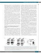
Figure 8. Accumulated NF-κB-inducing kinase (NIK) physically interacts with c-Abl and contributes to the NIK-c-Abl-STAT3 prosurvival complex formation. OPM-2 and RPMI-8226 cell lines were treated with MK-0457 at 0.4 μM and after 24 hours (h) of treatment were lysed and subjected to immunoprecipitation (IP) using (A) anti-NIK or (B) anti-c-Abl or control antibody (IgG) and immunoblotted (IB) with either NIK or c-Abl antibodies. Anti-c-Abl immunoprecititate filters were stripped and reprobed for STAT3. (C) Western blot of anti-NIK and anti-c-Abl immunoprecipitates results were subjected to densitometric scanning and protein expression under control conditions was set as 1. The histogram shows average quantification results±Standard Deviation (SD) of the association c-Abl/NIK from three immunopre- cipitations (*P<0.001 vs. untreated control cells, Dunnett and Tukey-Kramer tests).
haematologica | 2019; 104(12)
2475
