
Page 159 - 2019_12-Haematologica-web
P. 159
c-Abl/NIK limits the efficacy of Aurora inhibitors
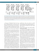
Figure 6. Aurora kinases inhibitors induce a cytoplasmic relocalization of c-Abl. Multiple myeloma (MM) cell lines were treated with MK-0457 (0.4 μM) and after 24 hours (h) cytoplasmic and nuclear extracts were prepared. Equal amount of Cytoplasmic (cyto) and nuclear (nuc) cell lysates (10 μg) were immunoblotted against c- Abl, phospho-c-Abl (Thr735), β Tubulin and Histone H3 as loading control of cytoplasmic and nuclear fraction, respectively. Bands were subjected to densitometric scanning: cytoplasmic and nuclear blots were normalized to total β Tubulin and Histone H3, respectively. The densitometric analysisis is reported in the graphs below: the relative fold change of cytoplasmic or nuclear c-Abl and phospho-c-Abl (Thr735) levels was normalized with respect to control condition, which was taken as 1. The ratio of cytoplasmic to nuclear c-Abl and phospho-c-Abl (Thr735) protein expression (Cyto/Nuc) is shown. The c-Abl and phospho-c-Abl (Thr735) Cyto/Nuc ratio in untreated cells was set as 1. In the histograms are shown average quantification results±Standard Deviation (SD) of four independent blots (°P<0.05, *P<0.005, **P<0.001 vs. untreated control cells, Dunnett test).
Both processes, Thr735 phosphorylation and concomi- tant cytoplasmic accumulation of c-Abl, were closely linked to NIK induction since its overexpression in MM cells increased Thr735 phosphorylation of cytoplasmic c- Abl (Figure 7A) and caused c-Abl to translocate from the nuclear to the cytoplasmic compartment, whereas its inhi- bition in these NIK-over-expressing cells reversed this shuttling (Figure 7A). These data were further confirmed by immunofluorescence analysis (Online Supplementary Figure S6).
A closer examination revealed that NIK was diffused in the cytoplasm with an accumulation around the nucleus of the tumor cells treated with pan-AKI (Figure 7B), and its overexpression also caused enhanced tyrosine phosphory- lation of cytoplasmic c-Abl (Figure 7A), to elicit its anti- apoptotic functions.15-18
Together with these results, siRNA-mediated knock- down of NIK completely abrogated the pan-AKI-induced Thr735 phosphorylation of c-Abl in OPM-2 and greatly decreased the high basal c-Abl Thr735 phosphorylation in the high NIK expressing JJN3 HMCL (Figure 7C).
Because pan-AKI can induce NIK accumulation and con- comitant c-Abl activation, and both these kinases con- verge on and activate the STAT3 pathway,10,17 we next investigated whether c-Abl can form a heterotrimeric complex with NIK and STAT3 in MM cells. As indicated in Figure 8A and B, there was little if any detectable inter- action of c-Abl and NIK in untreated MM cells. However, exposure of MM cells to Pan-AKI led to an increase in the association of c-Abl with NIK kinases that was at least a 3-fold higher than in untreated control cells (Figure 8C).
The interaction between NIK and c-Abl, and that previ- ously shown between NIK and STAT3 (Figure 4E), togeth- er with the fact that c-Abl can regulate the activation of STAT3 in cancer cells,17 indicated that these three proteins may form a trimeric complexes in pan-AKI-treated MM cells. Accordingly, as shown in Figure 8B, immunoprecip- itation of endogenous c-Abl from lysates of untreated or
pan-AKI-treated MM cells followed by STAT3 immunoblotting revealed that the pharmacological block- ade of Aurora kinases induced a physical interaction of c- Abl with STAT3, thus confirming that, in MM cells, pan- AKI can promote the formation of the ternary complex NIK-c-Abl-STAT3.
Pharmacological blockade of c-Abl sensitizes multiple myeloma cells to pan-AKI
To examine the functional significance of the pan-AKI- induced activation of c-Abl in MM cells we blocked its function using the Abl kinase inhibitors imatinib or nilo- tinib20 and monitored cell death in response to pan-AKI treatment. Both imatinib or nilotinib significantly increased the pan-AKI-induced cell death in the majority of the HMCL as well as in patient-derived primary MM cells, (P<0.005; n=9) (Figure 9A and B and Online Supplementary Figure S7A and B), with no significant differ- ences observed in the response rates of newly diagnosed (n=4) versus relapsed (n=5) patients (Online Supplementary Figure S8) and no effects seen in normal PBMC (Figure 9B and Online Supplementary Figure S7C).
In agreement with these results, Aurora-A and -B inhibi- tion by either Aurora A/B-specific siRNA or AMG-900,23,29 a potent and highly selective pan-AKI, significantly enhanced the sensitivity of MM cells to c-Abl inhibitors (Figure 9C, Online Supplementary Figures S9 and S10A and B). Furthermore, c-Abl kinase inhibitors consistently syn- ergized with pan-AKI to induce cell death in MM cells (Online Supplementary Figure S11 and Online Supplementary Table S3).
Remarkably, as observed in the majority of the HMCL analyzed, treatment of cells isolated from MM patients with Pan-AKI induced NIK accumulation, increased Thr735, Tyr245 and Tyr412 phosphorylation of c-Abl and Ser727 and Tyr705 phosphorylation of STAT3 (Figure 9D). None of these conditions was observed in similarly treat- ed PBMC from healthy donors.
haematologica | 2019; 104(12)
2473
