
Page 163 - 2019_12-Haematologica-web
P. 163
c-Abl/NIK limits the efficacy of Aurora inhibitors
can significantly contribute to the basal NF-κB activity of MM cells and that their inhibition can partially compen- sate for the NIK-induced activation of NF-κB pathways.
In MM, the pervasive DNA damage triggers constitutive activation of the ATR/ATM-regulated DNA damage response proapoptotic network which in turn leads to a
prominent and preferential nuclear localization of c-Abl. Here, however, it is unable to induce apoptosis because of disruption of the ABL-YAP1-p73 axis.21 The nuclear accu- mulation of c-Abl in MM21 may explain its marginal role in MM pathogenesis46 and the therapeutic inefficacy of c-Abl inhibitors in monotherapy regimens or when used in com-
A
BC
D
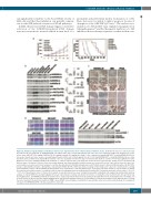
Figure 10. Pharmacological inhibition of Abl kinase improves the anti-myeloma effect of Aurora kinases inhibitors in vivo. (A) NOD-SCID mice were subcutaneously inoculated in the left flank with 107 RPMI-8226/R5 cells. When tumor size reached approximately 250 mm3, mice were randomly assigned (n=12/group) to receive vehicle alone, MK-0457 (50 mg/kg), PHA-680632 (50 mg/kg), imatinib (50 mg/kg twice daily), or the combination imatinib/MK-0457 or imatinib/PHA-680632 for two weeks. Results are tumor volume, mean±Standard Deviation (SD) mm3, plotted against time (P<0.001 imatinib/MK-0457 or imatinib/PHA-680632 vs. either treatment alone; Dunnett test). Kaplan-Meier survival curve was evaluated from the first day of treatment until death or sacrifice (P<0.0015, Log-Rank test after Bonferroni correction, imatinib/pan-AKI-treated animals vs. either treatment alone). The black bar on the abscissa represents the 14-day period of treatment. (B) After five days of treatment, four mice from each treatment group were humanely killed, and the tumors were removed for assay. Tumor tissues from mice were har- vested and processed for western blot analysis to monitor phospho-Aurora A (Thr288), phospho-Aurora B (Thr232), Aurora A, Aurora B, NIK, phospho-c-Abl (Thr735), phospho-c-Abl (Tyr245), phospho-c-Abl (Tyr412), c-Abl, phospho-STAT3 (Tyr705), STAT3 and Actin as loading control. Bands were subjected to densitometric scanning and normalized to Actin. c-Abl and STAT3 phosphorylations were normalized to c-Abl and STAT3 levels, respectively. The blots shown are representative of similar observations in four different mice receiving the same treatment. Histograms show mean±Standard Deviation (SD) of densitometry results from four mice (*P<0.01, **P<0.001, Tukey-Kramer test). (C) Tumors from vehicle or pan-AKI treated mice were formalin fixed paraffin embedded and analyzed by immunohistochemical analysis of c-Abl and NIK. The microphotographs shown are representative of similar observation in four mice receiving the same treatment (20x, 40x and 100x orig- inal magnification). (D) RPMI-8226/R5-derived tumors were analyzed by immunohistochemical staining for phospho-Histone H3, hematoxylin and eosin (H&E), and cleaved caspase-3 (4x, 10x and 20x original magnification). The microphotographs shown are representative of similar observations in four different mice receiving the same treatment. Western blot analysis for PARP, cleaved-PARP, cleaved caspase-3 and Actin of representative mice from each treatment group; for comparison, cell lysate from RPMI-8226/R5 cells was loaded in the same gel.
haematologica | 2019; 104(12)
2477
