
Page 155 - 2019_12-Haematologica-web
P. 155
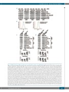
c-Abl/NIK limits the efficacy of Aurora inhibitors
A
B
CD
Figure 3. NF-κB-inducing kinase (NIK) accumulation interferes with inhibitory activities of pan-AKI NF-κB. (A) siRNA silencing of NIK but not the non-specific control siRNA, led to a decrease in NIK protein expression. Three hours (h) after electroporation, multiple myeloma (MM) cell lines were treated with the pan-AKI MK-0457 (0.4 μM) or PHA-680632 (1 μM). After 48 h, MM cell lines were lysed and subjected to western blot analysis to monitor the expression of NIK, phospho-IKKa/β, phospho-NF-κB2 p100, phospho-IκBa and Actin as loading control. All western blotting results were evaluated by densitometric scanning, corrected with respect to Actin expression, and expressed relative to the value obtained with the corresponding control set as 1. The relative protein amount is reported below the lanes. (B) RPMI-8226 and 8226/R5 were stably transfected with an empty vector or with plasmid expressing NIK protein. Pools of stable clones (8226-NIK and 8226/R5-NIK) were obtained by selection with puromycin. Both expression vectors co-expressed GFP to monitor the transfection by flow cytometry. Plots represent GFP fluorescence of cells transfected with empty vector (Blank) or that encoding for NIK compared to non-transfected cells. (C) NIK overexpression enhances nuclear localization and DNA transactivation activity of NF-κB subunits. Stable clones of RPMI-8226 and 8226/R5 transfected with empty vector (Blank) or expressing NIK protein or untrans- fected cells were seeded at a density of 2x10⁵ cells/mL. After 24 h cytoplasmic and nuclear extracts were prepared using the Active Motif's Nuclear Extract Kit. Cytoplasmic cell lysates were immunoblotted against NIK, p-IKKa/β, p-NF-κB2, p-IκB-a and tubulin as marker of cytoplasmic separation as well as loading control; nuclear extracts were immunoblotted against NF-κB p65, NF-κB1 p50, NF-κB2 p52, RelB and histone H2B as nuclear loading control; bands were subjected to den- sitometric scanning and the number below each lane represents the relative amount of the indicated proteins normalized to tubulin or histone H2B expression. Graphs below represent DNA binding activity of the NF-κB p65, NF-κB1 p50, NF-κB2 p52 and RelB subunit from the same nuclear extracts (TransAM NF-κB ELISA kit); results were normalized to the untransfected control (Untr). Values represent mean±Standard Deviation (SD) of three separate experiments. (**P<0.01 vs. untransfected condition; Dunnett’ test). (D) NIK inhibition attenuates NF-κB signaling. siRNA silencing of NIK but not the non-specific control siRNA, led to a decrease in NIK protein expression. RPMI-8226-NIK and 8226/R5-NIK were electroporated with control siRNA or with NIK siRNA. After 24-h cytoplasmic and nuclear extracts from transfected and untransfected cells were prepared. Cytoplasmic cell lysates were immunoblotted against NIK, phospho-IKKa/β, p-NF-κB2 p100, p-IκB-a and tubulin as marker of cytoplasmic separation as well as loading control; nuclear extracts were immunoblotted against NF-κB p65, NF-κB1 p50, NF-κB2 p52, RelB and Histone H2B as nuclear loading control; bands were subjected to densitometric scanning and the number below each lane represents the relative amount of the indicated proteins normalized to Tubulin or Histone H2B expression. Graphs below represent DNA binding activity of the NF-κB p65, NF-κB1 p50, NF-κB2 p52 and RelB subunit from the same nuclear extracts; results were normalized to the untransfected control (Untr). Values represent mean±SD of three separate experiments. (*P<0.05 vs. control siRNA condition; Dunnett test).
haematologica | 2019; 104(12)
2469
