
Page 143 - 2019_12-Haematologica-web
P. 143
Biomarkers of progression to multiple myeloma
(MCP-3), macrophage inflammatory protein-1 alpha (MIP- 1a), vascular endothelial growth factor (VEGF), fibroblast growth factor-2 (FGF-2), fractalkine, and transforming growth factor-alpha (TGF-a).12 However, the study by Vermeulen et al.12 that documented these associations did not allow investigation of changes in immune markers in relation to the risk of progression to full-blown disease.
Herein we aimed to replicate the inverse association between myeloma risk and blood levels of MCP-3, MIP- 1a, VEGF, FGF-2, fractalkine, and TGF-a observed by Vermeulen et al.12 We hypothesized that pre-diagnostic marker levels might be useful for predicting progression to MM. To this end, we analyzed MCP-3, MIP-1a, VEGF, FGF-2, fractalkine, TGF-a,12 and four additional markers that have been related to MM pathobiology – macrophage inflammatory protein-1 beta (MIP-1β),13 interleukin (IL)-13,14 tumor necrosis factor-alpha (TNF-a),15 and IL-1016 – in repeated pre-diagnostic plasma samples from 65 myeloma cases and 65 matched cancer-free controls. The utility of the candidate biomarkers in the prediction of the development of MM was evaluated by means of a multi- variable model including known risk factors for progres- sion.
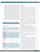
Table 1. Characteristics of the study population and the blood samples. Cases Controls Pa
Methods
Study population
The study was designed as a case-control study nested in a large population-based prospective cohort called the Northern Sweden Health and Disease Study (NSHDS).17 Within NSHDS, peripheral blood samples have been collected from the general population, with informed consent, since 1984. All collected samples are frozen within 1 h of the blood having been drawn and thereafter stored at -80°C at Umeå University Hospital (Sweden). At the time of sample selection for this study (October 2013), NSHDS contained samples from more than 100,000 individuals. Through linkage with the Swedish Cancer Registry, we identified incident myeloma cases (diagnosed between 1997 and 2013) who had pre- viously donated at least two pre-diagnostic blood samples within NSHDS (n=66). Cancer-free controls were selected from the same cohort, and were matched to cases, in a 1:1 ratio, for sex, age at blood sample collection (± 5 months), and date of blood sample collection (± 2 months) (Table 1).
Case classification was performed according to ICD-O-3.18 After acquisition of clinical data by retrospectively studying the patients’ records, one case was reclassified as MGUS, thus leaving 65 future myeloma cases for inclusion in the present study. The retrospective record review revealed that at the time of myeloma diagnosis, 43 cases had MM and 22 had SMM, based on the crite- ria of the International Myeloma Working Group (IMWG) from 2003.19 Twenty-five of the included cases were included in anoth- er study based on single samples per participant.12 This study was approved by the ethical review board at Umeå University (n. 08- 215M and 2017/242-31).
Immune marker and M-protein assessment
Ten immune markers were measured in duplicate in all samples (n=260) by a Luminex multiplex assay from Millipore (USA): MCP-3, MIP-1a, MIP-1β, VEGF, FGF-2, fractalkine, TGF-a, IL-13, TNF-a, and IL-10. Samples from matched cases and controls were included in random order in the same analytical batch. Laboratory personnel were blinded concerning case-control status and chronological order of samples. All analyses were performed according to the manufacturer’s protocol (Online Supplementary Methods).
M-proteins were assessed in samples from all future myeloma cases except four (due to insufficient sample volumes) by protein electrophoresis, immunofixation electrophoresis, and FLC assays (Online Supplementary Methods).
Statistical analyses
Immune marker concentrations were log10-transformed for nor- malization. Multiple imputation was applied to attain concentra- tion values when measurements were below the limit of quantifi-
20
The effect of immune marker levels on the probability of pro- gression to MM was evaluated by Kaplan-Meier plots and the log- rank test. For Kaplan-Meier estimates, time was calculated from repeated pre-diagnostic blood sample collection to either diagno- sis of a treatment-requiring condition or latest follow-up without signs of progression. To define cut-off values for immune marker concentrations between individuals progressing to MM and others without signs of progression, we performed receiver operating characteristic (ROC) analyses. Hazard ratio (HR) associations for risk factors of progression including dichotomized immune mark-
Age at sample collection, years Baseline sample, mean (range) Repeated sample, mean (range)
Sex Female Male
Body mass index
Baseline sample, mean (SD) Repeated sample, mean (SD)
Smoking status Baseline sample
Non-smoker Current smoker Former smoker
Repeated sample
Non-smoker Current smoker Former smoker
51 (30-69) 59 (40-74)
48 73.8
17 26.2
26.1 (3.8) 26.7 (3.7)
38 58.5 15 23.1 12 18.4
41 63.1 14 21.5 10 15.4
29 44.6 36 55.4
36 55.4 29 44.6
56 86.2 9 13.8
51 (30-68) 59 (40-74)
48 73.8
17 26.2
25.4 (4.0) 0.32 26.6 (4.0) 0.96
N%N%
65 65
Individual fasting status Baseline sample
40 61.5 14 21.5 11 17.0
42 64.6 12 18.5 11 16.9
32 49.2
33 51.8
36 55.4 29 44.6
57 87.7 8 12.3
0.94
0.90
0.60
1.00
0.80
0.50
≤ 8 hours > 8 hours
cation (3.4% of all data points).
trajectories between cases and controls were investigated by linear mixed models as described elsewhere,21 using the lme4 package in the R environment for statistical computing (The R Foundation for Statistical Computing) (Online Supplementary Methods).
Repeated sample ≤ 8 hours
> 8 hours
Thawing cycles before Baseline sample
No Once
Repeated sample
Differences in immune marker
No
Once 2 3.1 0
63 96.9
65 100.0
aP calculated using a paired t-test for continuous variables and the chi-square test for categori- cal variables. SD: standard deviation.
haematologica | 2019; 104(12)
2457
