
Page 89 - 2019_11 Resto del Mondo-web
P. 89
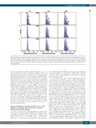
B. divergens infection in HbSS
A
B
C
Figure 3. Distribution of parasite sub-populations cultured in three types of red blood cells (RBC), based on genome content reveals all sub-populations are repre- sented in the cultures. (A) HbSS. (B) HbAS. (C) HbAA. Samples were collected at 1 hour (h), 24 h and 48 h and the percentage of each infected RBC sub-population was determined by FACS using VybrantRDyeCycleTMGreen dye to quantify the amount of parasitic DNA within infected cells, where (N) is the number of parasite genomes. (Top left) HbSS cells show higher numbers of 2N cells at 1 h time point compared to (left middle) HbAS and (left bottom) HbAA cells confirming a higher frequency of multiple invasion events in HbSS. (Middle and right panels) Parasite population structure at 24 and 48 h showing all sub-populations represented in all three cell genotypes.
ures” (2 attached parasites) and/or Maltese Cross (4 attached parasites) and/or double paired-figures (also as 4 parasites attached 2 by 2) and were seen co-existing with the detached ring parasites, showing that cellular division and differentiation of B. divergens followed the sequential transformation of stages from attached into unattached forms after completing cytokinesis before their egress. In addition, some of the infected RBC in HbSS cells hosted multiple parasites (≥4-8 or more) which were detected with variable frequency among HbSS cultures (Figure 4).
Thus, the morphological analysis supported the FACS analysis of population structures at the various sampled time points of the different cultures, HbAA, AS and SS. Importantly, the parasites in the HbSS RBC exhibited fea- tures that signaled they were ready for egress, i.e. the presence of both-infected RBC hosting multiple parasites as well as the presence of detached parasites in these cells. However, these parasites were not successful at starting new intra-erythrocytic cycles, as evidenced by the lack of increase in parasitemia.
Parasite infectivity is impaired in HbSS cells as the addition of fresh red blood cells does not rescue parasitemia in HbSS cultures
Success of the parasite culture propagation as measured by an increase in parasitemia requires successful invasion, successful production and maturation of merozoites which then need to successfully egress. This in turn depends on several factors involving host cells and para-
sites. Sickle cell anemia RBC are notorious for exhibiting increased fragility37 as compared to HbAA RBC and this could be a factor in the inability of the cultures to support parasitemia after 24 h.
To test whether the lack of increase in parasitemia was a consequence of defective egress, sickle cell fragility or defective merozoite maturation, we examined progres- sion of cultures in which the introduction of fresh RBC (either HbAA and HbSS) into 24 h HbSS parasite cultures was performed. After 1 h, all cells supported Babesia inva- sion equally well, as shown, following the mixing of merozoite inoculum and RBC (Online Supplementary Table S2). The cultures were then allowed to progress for 24 h at which point, the parasitemia in both the HbAA and HbSS cultures had risen slightly (HbAA-A: 3.2% vs. HbSS- A: 3.4%) (Online Supplementary Table S2), in line with the results reported for the 12 HbSS cultures above (Tables 1 and 2). The cultures were then split, either staying the same with only medium change (flasks A) or receiving fresh SS cells (flasks B) or AA cells (flasks C), maintaining hematocrits of 5%. At 48 h, the parasitemia in the original HbAA-A culture had doubled (from 3.2% to approx. 6.5%) while that of the original HbSS-A culture reported only a small increase in parasitemia (from 3.4% to approx. 4%), following the pattern reported for the original 12 HbSS cultures (Tables 1 and 2). The HbAA-B culture which was diluted with fresh HbAA RBC to receive half of the inoculum exhibited half the parasitemia of the undi- luted HbAA-A culture, once again as expected (3.2%)
haematologica | 2019; 104(11)
2195
