
Page 90 - 2019_11 Resto del Mondo-web
P. 90
J.R. Cursino-Santos et al.
(Online Supplementary Table S2). The two HbSS cultures that received fresh RBC, either HbAA (HbSS-C) or HbSS (HbSS-B), had lower parasitemias (2.1% and 2.5%, respectively; 24 h time point) than that of HbAA-B cul- ture, proving that the nature or condition of the host cells did not play a role in the inhibition of new cycles of para- sitemia seen in the HbSS cultures (HbSS-C and HbSS-B). At later time points (48 h after addition of fresh cells), this result of non-rescue was further strengthened when cul- tures in HBAA-B exhibited a robust increase in para- sitemia (16%), contrasting with HbSS-B and HbSS-C and the stable parasitemia of (3.6% and 2.3%, respectively). The original half culture which had only medium change (HbAA-A and HbSS-A) followed the previously reported inhibition patterns of in culture progression showing approximately 18% inhibition in HbSS-A culture growth, compared to the HbAA-A culture. Neither the addition of fresh HbSS RBC or HbAA RBC could significantly rescue the HbSS cultures, with the original HbSS-A parent cul- ture at 4.7% and the culture receiving fresh HbSS cells at 3.6% parasitemia. Surprisingly, the culture that received
fresh HbAA cells was even lower demonstrating an infec- tion rate of 2.3%, indicating that parasite development within the HbSS RBC was potentially impaired, resulting in the formation of merozoites unable to invade fresh RBC at frequencies typically seen in vitro parasite cultures. The formation of mature merozoites that egress from the RBC are critical to start new intra-erythrocytic cycles by invading new host cells, and these merozoites are required to be in an optimum invasive state to support new cycles.
Parasite egress from sickle red blood cells may also be compromised
One of the factors that may impact the progress in par- asitemia in HbSS cells is the ability of the parasite to egress from the host sickle cell and invade new host cells. The FACS profile of the parasite population in the HbSS cells was carefully examined to look for this inhibition of egress. The typical pattern of parasite-holding within the RBC that we have reported for B. divergens,31 when there is an egress defect, where the B. divergens parasite popula- tion builds its 4N and >4N populations to high levels that
ABCD
EFGH
IJLM
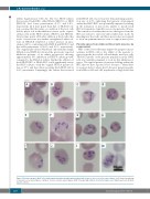
Figure 4. Parasites grown in HbSS cells exhibit atypical morphology although normal parasite forms are also seen in the same cultures. (A-C) Normal morphology of parasite seen as paired figures or Maltese Crosses or double paired figures (D-M) Unusually high numbers of detached rings seen at high frequency in all HbSS parasite cultures.
2196
haematologica | 2019; 104(11)
