
Page 77 - 2019_11 Resto del Mondo-web
P. 77
Deubiquitylase USP7 and human erythropoiesis
shown in Figure 4A, endogenous USP7 was immunopre- cipitated by anti-GATA1 antibodies but not by control IgG. Conversely, GATA1 was immunoprecipitated by anti-USP7 antibodies but not by control IgG (Figure 4B). To determine whether USP7 and GATA1 interact directly with each other, we performed GST pull-down assays under a cell-free condition by using purified recombinant GST-GATA1 and Flag-USP7 proteins. As shown in Figure 4C, the purified GST-GATA1 but not the control GST was able to pull down USP7. Furthermore, we mapped the detailed binding region of GATA1 and USP7 in
HEK293T cells, a non-erythroblast environment.38,42,43 Truncation mutants of GFP-USP7 and Flag-GATA1 were co-transfected into HEK293T cells and co-immunoprecip- itation analyses revealed that the N-terminal TRAF-like domain (1-208) of USP7 was critical for the interaction between GATA1 and USP7 (Figure 4D). Conversely, map- ping the region of GATA1 required for USP7 binding showed that the DNA binding domain (200-290) of GATA1 was responsible for its interaction with USP7 (Figure 4E). Collectively, these results show that USP7 interacts with GATA1 directly.
ABC
F
G
DE
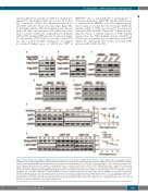
Figure 3. USP7 regulates the stability of GATA1. (A) Representative western blot analysis of HEK293T cells transfected with a GATA1-expressing plasmid and either an empty vector or increasing amounts of a GFP-USP7-expressing vector. (B) Representative western blot analysis of HEK293T cells that were transfected with a construct expressing Flag-GATA1 and an empty vector (-), a construct expressing wildtype USP7 (USP7-WT) or one expressing the USP7 (Cys223Ser) mutant (USP7- CS). (C) Representative western blot analysis of erythroblasts that were transfected with control (shNC) or USP7 shRNA treated with or without the proteasome inhibitor MG132 (20 mM, 6 h). (D) Representative western blot analysis of erythroblasts that were treated for 6 h with dimethylsulfoxide (DMSO) (-) and/or 15 mM P5091 (+) or with 15 mM P5091 and 20 mM MG132. (E) Representative western blot for the expression of GATA1 in erythroblasts treated for 6 h with DMSO (-) and/or 15 mM P22077 (+) or with 15 mM P22077 and 20 mM MG132 on day 9. (F) Erythroblasts transfected with control or USP7 shRNA were treated with cycloheximide (CHX) (150 mg/mL), and collected at the indicated times for western blot. Results are shown as mean ± standard deviation (SD) (**P<0.01). (G) Representative west- ern blot analysis of HEK293T cells that were transfected with a vector expressing Flag-GATA1 and an empty vector (NC), one expressing GFP-USP7-WT or one express- ing GFP-USP7-CS, after treatment with CHX (150 mg/mL) for the indicated amounts of time. Results are shown as mean ± standard deviation (**P<0.01). For all western blot analyses, GAPDH was used as the loading control.
haematologica | 2019; 104(11)
2183
