
Page 79 - 2019_11 Resto del Mondo-web
P. 79
Deubiquitylase USP7 and human erythropoiesis
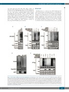
one lysine (K6, K11, K27, K29, K33, K48 or K63). As shown in Online Supplementary Figure S7, USP7 significant- ly decreased only the K48-linked poly-ubiquitin chain but not any other lysine isopeptide-linked poly-ubiquitin chains (K6, K11, K27, K29, K33 or K63). To further confirm that K48-linked poly-ubiquitin is removed by USP7, we replaced K48 or K63 lysine by arginine (R) and, as shown in Figure 5E, mutation of K48 but not K63 significantly impaired USP7-mediated deubiquitylation. Collectively, these results confirm that USP7 stabilizes GATA1 by removing the K48-linked poly-ubiquitin.
Discussion
Erythropoiesis is a process by which hematopoietic stem cells proliferate and differentiate to eventually pro- duce mature red blood cells. Many cellular and molecular changes occur during this process. Morphological changes include a progressive decrease in cell size, increase in chro- matin condensation and enucleation. At the molecular level, high-throughput analyses revealed dramatic changes in both the transcriptome and the proteome.2,44 In contrast to extensive studies on transcriptional networks, very little
ABC
DE
Figure 5. USP7 stabilizes GATA1 protein through deubiquitination. (A) Erythroblasts at day 7 transfected with control or USP7 shRNA (#1) lentivirus. GATA1 was immunoprecipitated with anti-GATA1 anyibody and immunoblotted with anti-ubiquitin on day 9. (B) Representative western blot analysis of ubiquitin after incubation of anti-Flag-coated beads with lysates from HEK293T cells that were transfected with empty vectors (-) or those expressing Flag-GATA1 either alone or in combination with vectors expressing USP7-WT or USP7-CS, and HA-ubiquitin. (C) Representative western blot analysis for ubiquitin after anti-Flag immunoprecipitation of HEK293T cells ectopically expressing Flag-GATA1 either alone or in combination with USP7-WT. Cells expressing both Flag-GATA1 and USP7-WT were treated with 20 mM P5091 or P22077 for 8 h before being harvested. (D) Representative western blot for the cell-free deubiquitylation assay. Ubiquitylated GATA1 was incubated with bacteri- al-expressed and purified USP7-WT for 2 h at 37°C, followed by western blot with anti-HA antibody (right panel). The left panel is the input. (E) Representative western blot analysis for ubiquitin after anti-Flag immunoprecipitation of HEK293T cells ectopically expressing Flag-GATA1 either alone or in combination with USP7-WT, and ubiquitin WT or mutant (K48R or K63R).
haematologica | 2019; 104(11)
2185
