
Page 75 - 2019_11 Resto del Mondo-web
P. 75
Deubiquitylase USP7 and human erythropoiesis
A
BC
DE
FG
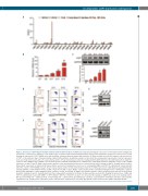
Figure 1. Deficiency of USP7 impairs human terminal erythroid differentiation. (A) RNA-sequencing data showing the expression of USP family members (fragments per kilobase of transcript per million) at each distinct stage of human terminal erythroid differentiation. (B) Real-time quantitative polymerase chain reaction results showing the expression of USP7 mRNA on the indicated days of human erythroid terminal differentiation. (C) Representative western blot analysis of the protein level of USP7 on the indicated days of human terminal erythroid differentiation. Quantitative analysis of data from three independent experiments of protein expression levels are shown (lower panel). (D) Left, representative profiles of flow cytometry-based detection of glycophorin A (GPA) expression in erythroblasts infected with the control or USP7 shRNA on day 9. Middle, representative profiles of Band3/α4-integrin levels of GPA-positive erythroblasts transfected with the control or USP7 shRNA lentviruses on days 11 and 13. Right, representative profiles of flow cytometry-based detection of enucleation by syto16 staining on day 13. Quantification from three independent experiments is indicated. (E) Representative western blot showing the protein level of γ-hemoglobin (HBG) and USP7 in erythroblasts transfected with either the control or USP7 shRNA on day 9. (F) Left, representative profiles of flow cytometry-based detection of GPA expression in erythroblasts treated with dimethyl- sulfoxide (DMSO) or USP7 inhibitor P5091 (5 mM) or P22077 (7.5 mM) on day 9. Middle, representative profiles of Band3/α4-integrin levels of GPA-positive erythrob- lasts treated with DMSO or USP7 inhibitor P5091 or P22077 on day 11 and day 13. Right, representative profiles of flow cytometry-based detection of enucleation by syto16 staining on day 13. Quantification from three independent experiments is shown. (G) Representative western blot showing the protein level of HBG and USP7 in erythroblasts treated with DMSO or USP7 inhibitors (P5091 or P22077) on day 9. For all western blot analyses, GAPDH was used as the loading control. BFU-E: burst-forming unit – erythroid; CFU-E: colony-forming unit – erythroid; ProE: proerythroblast; early Baso: early basophilic erythroblast; late Baso: late basophilic erythroblast; Poly: polychromatic erythroblast; Ortho: orthochromatic erythroblast; D: day; GPA: glycophorin A; FSC: forward scatter; DMSO: dimethylsulfoxide.
haematologica | 2019; 104(11)
2181
