
Page 66 - 2019_11 Resto del Mondo-web
P. 66
L. Shao et al.
immunoprecipitation assay showed reduction of Notch1 occupancy at the Notch-dependent Myc enhancer NDME37 and at Hes1, Hey1 and Dtx1 promoters38 in Notch1+/ΔTAD pBEC (Figure 6C). In line with our findings in thymocytes (Figure 2J), we observed a 3-fold accumulation of Notch1 ICN ΔTAD protein in pBEC when compared to cleaved Notch1 WT protein (Figure 5D). These results indicate that ΔTAD interference occurred in pBEC and contributed to the decreased target gene expression in EC, thus limiting regeneration and recovery of the BM niche.
For further in vitro study of ΔTAD transcriptional inter- ference, pBEC were sorted from the BM by expression of CD31+ and VE-cadherin+ (Online Supplementary Figure S8A). These cultured bone EC (cBEC) exhibited typical endothelial morphology, VE-cadherin+ adherent junc- tions and active Notch signaling (Online Supplementary Figure S8B, C). To replicate the effects of ΔTAD interfer- ence, we transduced cBEC with a retroviral vector expressing GFP (PMIGR1) or ICNΔTAD (Figure 5E, left). Accumulation of ICNΔTAD protein in GFP+ sorted cBEC
ABC
DE
FGHI
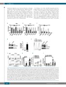
Figure 5. Notch1 ICN∆TAD interference in bone marrow endothelial cells. (A, B) The expression of the indicated genes was determined by quantitative reverse tran- scriptase polymerase chain reaction (RT-qPCR) in CD31+ primary bone endothelial cells (pBEC) at day 9 after treatment of wildtype (WT) and Notch1+/∆TAD littermates (n=3) with phosphate-buffered saline (PBS) or 5-fluorouracil (5-FU). Values were normalized to expression of genes in WT littermates. GAPDH was used as an internal expression control. (C) Local chromatin immunoprecipitation (CHIP) assay to detect the occupancy of Notch1 complex on Rbpj DNA-binding sites of Notch-dependent Myc enhancer (NDME), Hes1, Hey1, Dtx1 and GAPDH promoter elements. pBEC from WT and Notch1+/∆TAD mice were used for CHIP analysis at day 9 after 5-FU treat- ment (n=3). Values represent the mean of signal intensity relative to input DNA normalized to IgG. (D) pBEC from WT and Notch1+/∆TAD mice were harvested and used to measure protein levels of cleaved Notch1 and Notch1∆TAD by western blot (left panel). The band intensities of Notch1 ICN and Notch1 ICN∆TAD in Notch1+/∆TAD pBEC were quantified by ImageJ software (n=3, right panel). (E) Flow cytometry analysis of cultured bone marrow-derived endothelial cells (cBEC) (untransduced con- trol) or cBEC transduced with retrovirus expressing PMIGR1 or PMIGR1-ICN∆TAD (left panel). Western blot analysis for expression of Notch1 protein in transduced and sorted GFP+ cBEC expressing PMIGR1 or PMIGR1-ICN∆TAD. GAPDH was used as a loading control (n=3, right panel). (F, G) cBEC transduced with PMIGR1 or PMIGR1-ICN∆TAD were treated with 5-FU or vehicle (Veh) for 24 h (n=3). Representative annexin V and 4′,6-diamidino-2-phenylindole (DAPI) flow cytometry analysis (F) and quantified percentages of pro-apoptotic annexin V+ cells (G). (H) Cell counts of cBEC-PMIGR1 and cBEC-ICN∆TAD after Veh or 5-FU treatment. Cell numbers were counted at day 1 and day 2 after a single 5-FU treatment (n=3). (I) RT-qPCR for the indicated genes in cBEC-PMIGR1 and cBEC-ICN∆TAD cells 24 h after 5-FU treatment. Values were normalized to PMIGR1 transduced cells. GAPDH was used as an internal expression control. *P<0.05, **P<0.01, ***P<0.001.
2172
haematologica | 2019; 104(11)
