
Page 65 - 2019_11 Resto del Mondo-web
P. 65
Loss of Notch1 TAD interferes with niche recovery
and whether it affected the recovery of EC in the BM niche, we tested the expression of Notch target genes in pBEC. Under basal conditions, expression of Hes1, Hey1 and Dtx1 was downregulated in Notch1+/ΔTAD pBEC, while expression of Myc and EphB2 was unaffected (Figure 5A). However, 9 days after 5-FU treatment, expression of the
A
C
E
Notch target genes EphB2, Myc, Hes1, Hey1 and Dtx1 was markedly downregulated in Notch1+/ΔTAD pBEC (Figure 5B). To determine how Notch target expression was affected by the Notch1ΔTAD mutant, a chromatin immunoprecip- itation assay was performed on purified pBEC from WT or Notch1+/ΔTAD littermates after 5-FU treatment. The chromatin
B
F GHI
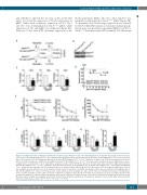
Figure 4. Conditional deletion of Notch1 in endothelial cells phenocopies Notch1+/∆TAD mice after myelosuppression. (A) Experimental design of 5-fluorouracil (5- FU) injection of Notch1f/f;VE-CadherinCreERT2+ mice. Tamoxifen (80 mg/kg) was injected intraperitoneally (IP) into Notch1f/f;VE-CadherinCreERT2+ mice for 5 consecutive days. One week later, 5-FU (150 mg/kg) was injected IP into Notch1f/f;VE-Cadherin CreERT2+ or CreERT2- littermates on days 1 and 14. (B) Protein expression of cleaved Notch1 in CD31+ primary bone endothelial cells (pBEC) from Notch1f/f;VE-Cadherin CreERT2- or CreERT2+ littermates after tamoxifen treatment. GAPDH was used as a loading control. (C) Quantitative reverse transcriptase polymerase chain reaction (RT-qPCR) expression of Hes1, Myc, Hey1 and Dtx1 in pBEC harvested from Notch1f/f;VE-Cadherin CreERT2+ or CreERT2- littermates after tamoxifen treatment. (D) Kaplan-Meier plot of Notch1f/f;VE-Cadherin CreERT2+ and Notch1f/f;VE-Cadherin CreERT2- littermates after tamoxifen and 5-FU treatment. Mice were monitored daily until day 28 after 5-FU injection (n=13-17 mice/group). Significance (P=0.05) was determined by the Mantel-Cox test. (E) Peripheral blood counts of red blood cells (RBC) and platelets (PLT), and concentration of hemoglobin (Hb) in Notch1f/f;VE-CadherinCreERT2+ and CreERT2- littermates were conducted before (D0) and at days 4, 7 and 9 after 5-FU injection. (F) Absolute numbers of hematopoietic stem cells (HSC) and Lin-Sca1+c-kit+ (LSK) cells in bone marrow were analyzed 9 days after the 5-FU injection and expressed as mean ± standard deviation (SD). (G) Absolute numbers of common lymphoid progenitors (CLP) in bone marrow were analyzed at day 9 after the 5-FU injection and expressed as mean ± SD. (H) Absolute numbers of pBEC were analyzed at day 9 after the 5-FU injection by flow cytometry. (I) Percentages of annexin V+ Lin-Sca1+c-Kit+ pBEC 9 days after 5-FU injection are presented as mean ± SD. *P<0.05, **P<0.01, ***P<0.001.
D
haematologica | 2019; 104(11)
2171
