
Page 64 - 2019_11 Resto del Mondo-web
P. 64
L. Shao et al.
decrease in Notch target expression (Figure 4C) in pBEC. The condition of Notch1f/fVE-cadherin-CreERT2+ mice deterio- rated (body condition index = 2) within a week after treat- ment, and five of 13 mice succumbed between days 7-9 after treatment (Figure 4D). The morbidity of Notch1f/fVE- cadherin-CreERT2+ mice was reflected in the low peripheral blood counts (Figure 4E) as well as decreases in the num- bers of BM HSC, progenitors and CLP (Figure 4F, G). Consistent with increased endothelial apoptosis after treat- ment, the absolute number of pBEC in Notch1f/fVE-cadherin- CreERT2+ mice was significantly reduced (Figure 4H, I).
Besides the endothelium, perivascular cells have been implicated in the regulation of HSC function.36 We observed a decrease in numbers of BM PDGFRβ+ pericytes after chemotherapy in Notch1+/ΔTAD mice. However, this effect was not due to increased apoptosis of the pericytes and may be attributed to a previously described loss of
ABC
BM EC (Online Supplementary Figure S7A). To determine whether Notch signaling in pericytes played a role in the recovery of the BM niche, RBPJf/f-PDGFRβ-CreERT2+ and RBPJf/+-PDGFRβ-CreERT2+ littermates were treated first with tamoxifen and then with 5-FU (Online Supplementary Figure S7B, C). We observed no significant hematopoietic differ- ences between the two cohorts after 5-FU treatment (Online Supplementary Figure S7D-I). Overall these results show that robust Notch signaling in EC is essential for recovery of the BM niche following chemotherapeutic challenge.
Loss of the transcriptional activation domain suppresses transcriptional activation of Notch1 targets in endothelial cells
To determine whether the ΔTAD-dependent transcrip- tional interference observed in T cells also occurred EC
DE
GHI
F
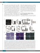
Figure 3. Defective recovery of endothelial niche induced by Notch1+/ΔTAD. (A) Whole mount imaging of the sternal vasculature after staining for CD31 and VE-cad- herin was performed in WT and Notch1+/∆TAD mice under resting conditions, including analysis of vessel length and vessel numbers (n=4). Scale bar=150 mm. (B) CD31+ primary bone endothelial cells (pBEC) were isolated at day 3 after 5-fluorouracil (5-FU) injection and expression of the indicated genes was determined by quantitative reverse transciptase polymerase chain reaction (RT-qPCR) normalized to expression of genes after injection of phosphate-buffered saline (PBS) (n=3). GAPDH was used as an internal expression control. Levels of active Notch1 protein in pBEC were measured by western blot at day 3 after 5-FU injection (insert). (C) Experimental design of 5-FU treatment for endothelial cell analysis in bone marrow (n=6). (D) pBEC harvested from long bones of WT and Notch1+/∆TAD littermates gated for 4′,6-diamidino-2-phenylindole (DAPI)-CD45-TER119-CD31+ and analyzed by flow cytometry for annexin V at day 9 after PBS or 5-FU injection (left panel). Percentages of annexin V+ cells in CD31+ endothelial cells (pBEC) are presented as mean± SD (right panel). (E) Isolated pBEC from digested bones were stained with DAPI-CD45-TER119-CD31+. The purity of CD31+ cells as a percent of CD31+CD45+ cells is demonstrated in the histogram analysis (left panel). The expression of indicated genes was determined by RT-qPCR in pBEC isolated from WT and Notch1+/∆TAD mice at day 7 after 5-FU injection (right panel). Fold changes in the relative gene expression, normalized to WT samples with GAPDH used as an internal expression control, are shown as mean±SD. (F) Absolute numbers of pBEC (DAPI-CD45- TER119-CD31+) were analyzed at day 9 after 5-FU injection by flow cytometry. (G) Representative longitudinal bone sections from mice treated with PBS (upper pan- els) or 5-FU (lower panels) stained with hematoxylin and eosin-Y at day 9 after 5-FU treatment. Scale bar=50 μm. (H, I) Representative longitudinal bone section from mice treated with PBS (upper panels) or 5-FU (lower panel) fixed with formalin and stained for CD31 (H, red, scale bar=20 mm) or endomucin (endmc) (I, green, scale bar=100 μm) at day 9 after 5-FU injection. DAPI was used to stain nuclei (blue). Filled white arrows point to vascular structures representative of the bone marrow endothelium. The open white arrow points to discontinuous vascular structures. **P<0.01, ***P<0.001.
2170
haematologica | 2019; 104(11)
