
Page 127 - 2019_11 Resto del Mondo-web
P. 127
CUDC-907 monotherapy in AML
induction of apoptosis begins at 12 h for MOLM-13 cells and 16 h for U937 cells (Figure 4C, D). Taken together, these results suggest that these changes in protein levels coincide with the induction of apoptosis.
Mcl-1 and Bim play important roles in CUDC-907- induced apoptosis
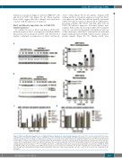
To confirm the roles of Mcl-1 and Bim in CUDC-907- induced apoptosis, Mcl-1 overexpression and Bim shRNA knockdown were performed in U937 cells. Western blot analysis confirmed overexpression of Mcl-1 and knock-
A
down of Bim (Figure 5A, B; left panels). Annexin V/PI staining and flow cytometry analysis revealed that Mcl-1 overexpression and Bim knockdown partially prevented CUDC-907-induced apoptosis (Figure 5A, B; right panels), providing evidence that Mcl-1 and Bim play roles in CUDC-907-induced apoptosis. Real-time reverse tran- scriptase PCR results showed that CUDC-907 treatment caused a concentration-dependent and significant increase of Bim transcripts, while Mcl-1 transcript levels remained largely unchanged in both AML cell lines and two primary patient samples (Figure 5C, D). Similar results were
B
CD
Figure 5. Mcl-1 and Bim play important roles in CUDC-907-induced apoptosis in acute myeloid leukemia cells. (A, B) U937 cells were infected with Precision LentiORF Mcl-1 (U937/Mcl-1) and RFP control (U937/RFP) (A) or NTC- (U937/NTC) and Bim-shRNA (U937/Bim) (B) lentivirus particles overnight, then washed and incubated for 48 h prior to the addition of blasticidin or puromycin, respectively, to the culture medium. The antibiotic-resistant cells were treated with CUDC-907 for 24 h. Whole cell lysates were subjected to western blotting. The fold changes for the Mcl-1 or Bim densitometry measurements, normalized to β-actin and then compared to no drug treatment control, are indicated (left panel). The cells were treated with CUDC-907 for 24 h and then subjected to annexin V/propidium iodide staining and flow cytometry analysis. ***P<0.001 (right panel). Bim S, L, and EL indicate Bim short, long, and extra-long isoforms, respectively. (C, D) MV4-11, U937 and MOLM-13 AML cell lines and two primary AML patient samples were treated with 0-100 nM CUDC-907 for 24 h. Total RNA was isolated and Bim (C) and Mcl-1 (D) transcripts were determined by real-time reverse transcriptase polymerase chain reaction (RT-PCR). *P<0.05, **P<0.01, and ***P<0.001. (continued on the next page)
haematologica | 2019; 104(11)
2233
