
Page 125 - 2019_11 Resto del Mondo-web
P. 125
CUDC-907 monotherapy in AML
D
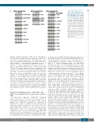
Figure 3. CUDC-907 treatment inac- tivates PI3K and ERK and causes downregulation of Mcl-1, CHK1, Wee1, and RRM1, and upregula- tion of Bim and γH2AX. (continued from the previous page) (D) NSG mice were injected with MV4-11 cells (1x10⁷ cells/mouse). After 21 days, the mice were randomized into three groups and treated with the vehicle control or a single dose of CUDC-907. 24 h after treatment, the mice were sacrificed and bone marrow cells were harvested. Human cells were enriched as described in the Methods section. Whole cell lysates were subjected to western blotting. Normalized densit- ometry measurements are shown below the corresponding blot. CUDC: CUDC-907.
internal tandem duplication (ITD) (n=14, median IC50 143.3 nM) were significantly more sensitive to CUDC-907 than those from patients without FLT3-ITD (n=61, medi- an IC50 217.6 nM; P=0.0281) (Figure 2A). CUDC-907 treat- ment induced a concentration-dependent increase of annexin V+ cells accompanied by increased cleaved cas- pase 3 and PARP (Figure 2B-D), demonstrating that CUDC-907 treatment induced apoptosis in primary AML samples ex vivo. Next, we treated five primary AML sam- ples with or without 100 nM CUDC-907 for 24 h and then plated the cells in methylcellulose. After 2 weeks, the number of surviving AML cells capable of generating leukemia colonies (AML-CFU) were enumerated. CUDC- 907 treatment significantly reduced the number of AML- CFU in all samples tested, indicating that CUDC-907 treatment decreased leukemia progenitor cells (Figure 2E). In contrast, CUDC-907 treatment did not have a signifi- cant effect on colony formation of normal bone marrow mononuclear cells (Figure 2F, G), suggesting that CUDC- 907 treatment spares normal hematopoietic progenitor cells.
CUDC-907 downregulates Mcl-1, CHK1, Wee1, and RRM1, and upregulates Bim in acute myeloid leukemia cells
As previously reported,15 CUDC-907 treatment decreased the levels of p-AKT (both T308 and S473) in three AML cell lines and two primary AML samples (Figure 3A). CUDC-907 treatment also decreased p- ERK1/2 levels, while total ERK levels remained relatively unchanged. p-AKT changes were detected as early as 3 h after CUDC-907 treatment in the cell lines (Online Supplementary Figure S1). These results confirm that CUDC-907 inactivated the PI3K/AKT and MEK/ERK pathways at these concentrations. Total AKT levels were decreased after 24 h of CUDC-907 treatment, although 3 h of treatment caused a decrease of p-AKT in the absence
of changes in total AKT (Online Supplementary Figure S1). Increased acetylation of histone H4 and tubulin (deacety- lated by HDAC6) was detected in the AML cell lines, con- firming inhibition of HDAC at these concentrations as early as 3 h after treatment (Figure 3A and Online Supplementary Figure S1). Substantially increased acetyla- tion of histone H4 was also detected in both primary AML samples, while the acetylation of tubulin was increased to a much lesser extent. Inhibition of the PI3K pathway and HDAC have been shown to cause downregulation of Mcl- 1 and upregulation of Bim, respectively.22-25 Accordingly, CUDC-907 treatment caused a reduction of Mcl-1 and an increase of Bim (Figure 3B), while Bcl-2, Bcl-xL, Bax, and Bak protein levels remained unchanged (Online Supplementary Figure S2). Based on the reports that HDAC inhibitors can downregulate DNA damage response pro- teins,23-26 we looked at γH2AX (a potential biomarker of DNA double-strand breaks), DNA damage response pro- teins CHK1, Wee1, and related downstream proteins. CUDC-907 treatment caused an increase of γH2AX and decreases of CHK1, p-CDK1, p-CDK2, Wee1, and RRM1 in AML cell lines and primary AML samples (Figure 3C). Total CDK1 and CDK2 levels were largely unaffected. p- CDC25C and RRM2 decreased, except in U937 cells. E2F1 levels decreased in the cell lines and in one primary AML samples. These results suggest that CUDC-907 may induce DNA damage, which causes death of AML cells. The above results were further confirmed in the MV4-11 xenograft mouse model following administration of a sin- gle dose of CUDC-907 (Figure 3D). Downregulation of CHK1, Wee1, and RRM1 by CUDC-907 treatment was not affected by the pan-caspase inhibitor Z-VAD-FMK (Online Supplementary Figure S3). In contrast, Z-VAD-FMK treatment itself caused an increase in Mcl-1 protein levels, suggesting that caspases are involved in the regulation of Mcl-1 protein. Surprisingly, co-treatment of AML cells with CUDC-907 and Z-VAD-FMK resulted in substantial-
haematologica | 2019; 104(11)
2231
