
Page 128 - 2019_11 Resto del Mondo-web
P. 128
X. Li et al.
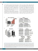
obtained in MV4-11 xenograft samples following a single dose of CUDC-907 (Figure 5E). A Mcl-1 protein stability assay using cycloheximide (10 mg/mL) revealed that Mcl-1 levels decreased faster in CUDC-907-treated cells than in vehicle-treated cells, resulting in a significantly shorter half-life (MOLM-13: 58 vs. 83 min, P=0.0266; U937: 79 vs. 94 min, P=0.0211) (Figure 5F, G). These results demon- strate that CUDC-907 downregulates Mcl-1 expression by decreasing the stability of Mcl-1 protein.
Phosphorylation of Mcl-1 at T163 has been shown to stabilize Mcl-1 by prolonging its half-life27 and phosphory- lation at S159 enhances ubiquitylation and degradation.28 In MOLM-13 and U937 cells, CUDC-907 treatment caused downregulation of p-Mcl-1 (T163), while p-Mcl-1
EF
(S159) levels remained unchanged (Figure 5H; upper panel). Treatment with the proteasome inhibitor MG-132, prevented downregulation of Mcl-1 by CUDC-907 (Figure 5H; lower panel). Since ERK has been reported to phos- phorylate Mcl-1 at T16327 and CUDC-907 treatment inac- tivates ERK, we treated MOLM-13 cells with the ERK inhibitor SCH-772984 and found that treatment did indeed downregulate p-Mcl-1 (T163), while having little to no effect on p-Mcl-1 (S159) levels (Online Supplementary Figure S6A). MG-132 treatment prevented downregulation of Mcl-1 following SCH-772984 treatment (Online Supplementary Figure S6B). Taken together, these results suggest that CUDC-907 inactivates ERK, resulting in decreased Mcl-1 stability and Mcl-1 protein levels.
GH
Figure 5. (continued from the previous page) (E) Cells obtained from the MV4-11 xenografts, which were treated with a single dose of CUDC-907, were enriched for human cells. Then total RNA was isolated and real-time RT-PCR performed to determine Mcl-1 and Bim transcripts. ***P<0.001. (F, G) MOLM-13 and U937 cells were treated with vehicle control, 50 nM CUDC-907 or 100 nM CUDC-907 for 12 h, washed and then treated with cycloheximide (CHX) for up to 2 h. Whole cell lysates were subjected to western blotting and probed with anti-Mcl-1 or anti-β-actin antibody. The fold changes for the Mcl-1 densitometry measurements, normalized to β- actin and then compared with no drug treatment control, are shown as mean ± standard error of mean. *P<0.05. (H) MOLM-13 and U937 cells were treated with CUDC-907, MG-132, or MG-132 plus CUDC-907 for 24 h. Western blot analyses of whole cell lysates are shown. The fold changes for the densitometry measure- ments, normalized to β-actin and then compared to no drug treatment control, are indicated. RFP: red fluorescent protein; CUDC: CUDC-907; NTC: non-treated control; AML: acute myeloid leukemia; MG: MG-132, a proteasome inhibitor.
2234
haematologica | 2019; 104(11)
