
Page 200 - 2019_10 resto del Mondo_web
P. 200
E. Karampini et al.
lular protein trafficking as well as the efficiency of WPB to exocytose upon stimulation in two clonal VAMP8-/- BOEC lines. To assess the involvement of VAMP8 in a fusion step between WPB and the endosomal compartment during CD63 recruitment, we checked the localization of CD63 in control and VAMP8-deficient cell lines. Immunofluorescent staining of CD63 and vWF in the VAMP8-/- lines exhibit a similar pattern when compared to the control cell lines, with CD63 being found on endo- some-like structures as well as WPB (Online Supplementary Figure S6B). This suggests that membrane transfer between endosomes and WPB during CD63 trafficking does not depend on VAMP8. We next examined the involvement of VAMP8 in WPB exocytosis by testing stimulus-induced vWF release in CRISPR-edited VAMP8-/- BOEC. Our data show that the intracellular levels of vWF are similar for VAMP8-deficient and control cell lines (Figure 4C), show- ing that the process of CRISPR-Cas9 genetic modification and clonal selection does not affect vWF biosynthesis and/or WPB biogenesis. However, upon Ca2+-mediated stimulation of WPB release with histamine, VAMP8 knock- out endothelial cells secreted significantly less vWF when compared to the control lines (Figure 4D). These findings demonstrate that VAMP8 promotes stimulus-induced vWF secretion and establish VAMP8 as a novel component of the WPB exocytotic machinery.
A
Discussion
The AP-3 complex regulates the formation and matura- tion of lysosome-related organelles in many different cell types.8 In this study, we show that mutations in the AP3B1 gene that lead to HPS-2 result in loss of AP-3 β1 and rapid degradation of the AP-3 μ1 subunit of the AP-3 complex. While the δ1 and s1 subunits are still expressed, the con- sequential destabilization of the AP-3 complex leads to a failure to traffic proteins to the WPB. Two of these, CD63 and VAMP8, normally co-reside on endosomes and WPB. However, in the absence of AP-3-dependent sorting, their fates differ radically. Blockade of the route from the endo- somal compartment to the WPB causes an increase of CD63 on the cell surface, possibly caused by a global redistribution of CD63 in the absence of its storage com- partment. This has previously been reported for other cell types derived from HPS-2 patients, such as cytotoxic T lymphocytes and fibroblasts.13-15,23 VAMP8 expression is severely reduced in both HPS-2 BOEC and in CRISPR- engineered AP3B1 KO BOEC, for which we currently do not have an explanation. Contrary to CD63, VAMP8 does not contain any of the known sorting motifs that would allow it to directly interact with the AP-3 complex.9,10 VAMP7, a v-SNARE that is found on a number of LRO, has been shown to interact with the AP-3 complex on
B
CD
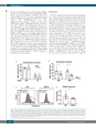
Figure 3. Impaired stimulus-induced von Willebrand Factor (vWF) secretion and P-selectin cell surface exposure in HPS-2 blood outgrowth endothelial cells (BOEC).
(A) Basal (unstimulated) secretion of vWF in conditioned media after 24 hours and 1 hour from WT HPS-2 (black) and HPS-2 (white) BOEC (n=6). (B) Stimulated vWF secretion after 30 minutes treatment with 100 μM histamine (HIS) or 10 μM forskolin + 100 μM IBMX (FSK) from WT (black) and HPS-2 (white) BOEC. Secretion is expressed as relative proportion of intracellular vWF in unstimulated cells (n=9). (C) Representative histograms of P-selectin (CD62P) cell surface expression before (close gray) and after 5 minutes stimulation with 100 μM histamine (open black line) (D) CD62P exposure after histamine stimulation expressed as fold increase over unstimulated cells (n=8). Two tailed Student t-test, *P<0.05.
2096
haematologica | 2019; 104(10)
