
Page 201 - 2019_10 resto del Mondo_web
P. 201
AP-3 dependent trafficking of VAMP8 in endothelial cells
early endosomes via a direct interaction between the AP-3 δ1 subunit and the N-terminal longin domain of VAMP7.26,27 Destabilization of the AP-3 complex, such as in mocha mice which lack the δ1 subunit of AP-3, leads to mistargeting of VAMP7.27 It has also been suggested that AP-3 indirectly traffics STX13 to maturing melanosomes via VAMP7.28 Although VAMP8 does not contain a longin domain, it may be possible that during WPB maturation there is a similar mechanism by which VAMP8 latches on to the back of another AP-3 interacting partner.
In this study, we further explored the effect of WPB maturation on regulated secretion and identify VAMP8 as a novel component of the exocytotic fusion machinery. Previous studies have found that WPB contain the v- SNAREs VAMP3 and VAMP8, but so far only VAMP3 has been implicated in Ca2+-dependent WPB exocytosis.25 We had previously established that VAMP8 is a direct interac- tor of both syntaxin-3 (STX3) and syntaxin-4 (STX4).21 STX4, which is found at the plasma membrane, has been implicated in thrombin-induced release of WPB through the formation of a complex with WPB-localized
VAMP3.25,29 We have recently shown that STX3 is local- ized on WPB and that absence of STX3 from endothelial cells results in impaired basal and hormone-evoked vWF secretion.21 The decrease in histamine-evoked vWF secre- tion in VAMP8 knock-out endothelial cells indicates an active involvement of VAMP8 in stimulus-induced WPB exocytosis. Based on its ability to interact with STX4 and STX3, VAMP8 can potentially support exocytosis via direct (WPB-plasma membrane) or homotypic (WPB- WPB) fusion modes. Further research on this topic should address the involvement of VAMP8 in the different modes of fusion.
In vivo, VAMP8-/- mice show delayed and decreased thrombus formation.30 Although, in their study, Graham et al. found that defective thrombus formation correlated with impaired dense granule release from VAMP8-/- platelets, they did not test whether lack of VAMP8 in endothelial cells also contributed to defects in thrombus formation. A more pronounced defect in thrombus forma- tion was also observed in ruby eye mice, a murine model for HPS-6, which lack platelet dense granules.31
AB
CD
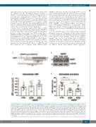
Figure 4. CRISPR-Cas9-engineered VAMP8-/- blood outgrowth endothelial cells (BOEC) have decreased histamine-induced von Willebrand Factor (vWF) secretion. A and B) Generation of clonal VAMP8 deficient BOEC lines using CRISPR/Cas9 genome-editing. (A) Cartoon representation of the CRISPR/Cas9-engineering strategy to generate VAMP8-/- BOEC lines. Guide RNA targeting exon 1 and exon 2 (gRNA3 and gRNA4&5, respectively) are shown underneath fragments of VAMP8 exon 1 and 2 sequences with their protospacer adjacent motif (PAM) indicated in red. (B) Loss of VAMP8 expression in clonal VAMP8-/- BOEC. Lysates of clonal CTRL BOEC and 2 clonal VAMP8-/- BOEC lines (34.B4 and 35.B1) were separated with SDS-PAGE and were immunoblotted for VAMP8; α-tubulin was used as a loading control. Molecular weight standards are indicated on the left (kDa). (C) Intracellular levels of vWF in lysates from control and 2 clonal VAMP8-/- KO BOEC lines (34.B4 and 35.B1) as determined by ELISA (n=9). (D) Stimulated VWF secretion after 30 minutes treatment with 100 μM histamine (HIS) from CTRL (close black) and 2 clonal VAMP8-/- KO BOEC lines (34.B4 and 35.B1) (open circle and square respectively). Secretion of vWF in supernatant from control and 2 clonal VAMP8-/- KO BOEC lines (34.B4 and 35.B1). Secretion is expressed as relative proportion of intracellular vWF in unstimulated cells (n=9). Two tailed Student t-test, ns: P>0.05; *P<0.05; ***P<0.001.
haematologica | 2019; 104(10)
2097
