
Page 199 - 2019_10 resto del Mondo_web
P. 199
AP-3 dependent trafficking of VAMP8 in endothelial cells
Weibel-Palade bodies v-SNARE protein VAMP8 promotes Weibel-Palade body exocytosis
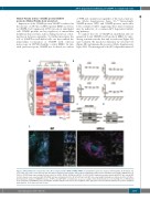
Expression of the SNARE protein VAMP8 is reduced in the absence of AP-3 β1, in HPS-2 patient BOEC as well as in CRISPR-Cas9 engineered AP3B1 knockout endothelial cells. SNARE proteins are key regulators of intracellular membrane fusion events, such as during exocytosis or dur- ing fusion between organelles. To further investigate the role of VAMP8 in endothelial cells, we first studied the intracellular localization of VAMP8 with confocal microscopy in HUVEC/healthy control BOEC. In line with previous reports, VAMP8 was localized on a subset
AB
of WPB and on spherical organelles of the endosomal sys- tem (Online Supplementary Figure S5).21,25 Interestingly, VAMP8-positive WPB and VAMP8-positive endosomes both contained CD63, suggesting their shared itinerary may be indicative of a common AP-3 dependent traffick- ing pathway.
To explore the role of VAMP8 in endothelial cells we generated clonal CRISPR-Cas9 knockout BOEC by intro- ducing a mutation in the first and second exon (Figure 4A) and evaluated the knockout efficiency by western blot (Figure 4B) and immunofluorescence (Online Supplementary Figure S6A). We investigated both AP-3-dependent intracel-
E
Figure 2. HPS-2 BOEC lack components of the AP-3 complex and the WPB v-SNARE VAMP8. (A and B) Whole proteome analysis of HPS-2 BOEC. (A) Heatmap of Z- scored LFQ (log2) values of the proteins with the highest variation between BOEC derived from 4 individual healthy donors and HPS2 derived BOEC (ANOVA S0=0.4, FDR = 0.05) (B) Graph representing LFQ (log2) values for AP3B1, AP3D1, AP3M1 and AP3S1. (C) HPS-2 BOEC lentivirally transduced with mEGFP-AP-3β1 and mEGFP (control). Lysates were separated with SDS-PAGE and were immunoblotted for AP-3β1, AP-3μ1 and GFP; molecular weight standards are indicated on the left (kDa). (D) Immunoblot analysis of VAMP8 in lysates of HPS-2 and WT BOEC; α-tubulin was used as a loading control.(E) Immunofluorescent stainings for von Willebrand factor (magenta) and VAMP8 (cyan) in healthy WT and HPS-2 BOEC. Boxed areas are magnified in the right part. Yellow arrowheads indicate the position of WPB in both channels. Scale bars represent 10 μm.
CD
haematologica | 2019; 104(10)
2095
