
Page 153 - 2019_10 resto del Mondo_web
P. 153
p66shc deletion exacerbates leukemia in TCL1 mice
Consistent with our finding that the upregulation of sur- face CXCR4 in CLL cells is mainly controlled post-transla- tionally,11 no correlation was observed between the mRNA levels of p66Shc and CXCR4 (Figure 6E and Online Supplementary Figure S10C).
The analysis was extended to CCR2 and CXCR3, which were selectively overexpressed in UM-CLL cells (Online Supplementary Figure S9C,D). Similar to CCR7, expression of these receptors was inversely correlated with that of p66Shc (Figure 6F,G and Online Supplementary Figure S10D,E), suggesting that p66Shc may negatively modulate their expression. p66Shc reconstitution in CLL cells did indeed result in a decrease in CCR2 and CXCR3 mRNA (Figure 6H and Online Supplementary Figure S10F).
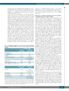
Interestingly, infiltration of both nodal and extranodal areas, assessed by the number and size (cm) of infiltrated lymph nodes and the presence of spleen and/or liver enlargement, was significantly greater in patients whose leukemic cells had p66Shc mRNA levels below an arbi- trarily set threshold (0.24, corresponding to the mean DDCt p66Shc mRNA of all CLL patients) (Online Supplementary Figure S11 and Table 1). These data provide evidence of a correlation of the severity of the p66Shc expression defect in CLL cells with their ability to infil- trate both nodal and extranodal districts, strongly sup- porting a role for p66Shc deficiency in disease presenta- tion. Of note, p66Shc expression was enhanced in CLL patients showing a significant response to second-line ibrutinib treatment but not in CLL patients who failed to
Table 1. Pathological characteristics of patients with chronic lymphocytic leukemia and p66Shc mRNA levels in their respective leukemic cells during
respond to ibrutinib therapy (Table 1 and Online Supplementary Table S1), suggesting that the response of CLL patients to therapeutic regimens results, at least in part, from the ability of leukemic cells to restore p66Shc expression.
Modulation of CCR2 and CXCR3 expression by p66Shc is mediated by its pro-oxidant activity
p66Shc has a ROS-elevating activity that depends on its ability to interact with cytochrome c and interrupt the respiratory chain.4 We quantified homeostatic ROS pro- duction in CLL cells loaded with the cell-permeant probe CM-H2DCFDA. ROS production was profoundly decreased in CLL B cells compared to that in normal B cells, with the lowest levels in UM-CLL patients (Figure 7A), consistent with their lowest p66Shc levels.6 Furthermore, we found a direct correlation between ROS production and p66Shc expression in CLL cells (Figure 7B). These findings were recapitulated in CM-H2DCFDA- loaded Eμ-TCL1 cells which, similar to CLL cells, express low levels of p66Shc (Figure 1B) and in which ROS pro- duction was lower than that in B cells from control C57BL/6 mice (Figure 7C). ROS production was further impaired in Eμ-TCL1/p66Shc-/- cells (Figure 7C), confirm- ing the pro-oxidant activity of p66Shc.
Transcription of both ccr7 and s1pr1 is controlled in opposite directions by the ROS-elevating activity of p66Shc.8 To address the potential role of the pro-oxidant function of p66Shc in the regulation of CCR2 and CXCR3 expression we used the CLL-derived human B-cell line MEC1 stably transfected with a ROS-defective mutant carrying a E→Q substitution at positions 132-133 (p66QQ), which disrupts cytochrome c binding (Figure 7D,E).8 The empty vector transfectant lacking p66Shc (ctr) and a transfectant expressing the wildtype protein (p66) were used as controls. Flow cytometric analysis of home- ostatic ROS production in the CM-H2DCFDA-loaded MEC1 transfectants showed enhanced ROS production in p66Shc-expressing cells, but not in cells expressing p66ShcQQ, compared to control cells (Figure 7F).
Mitochondrial redox signaling and apoptosis are also modulated by p53,36 which is mutated in a large propor- tion of CLL patients1 as well as in MEC1 cells.37 To rule out a role for TP53 mutations in the enhanced ROS pro- duction by p66Shc-expressing MEC1 cells, ROS were measured in EBV-immortalized B cells, which express wildtype p53,38 transiently depleted of p66Shc by short interfering RNA-mediated knock-down. Similar to MEC1 cells, p66Shc deficiency in EBV-immortalized B cells resulted in a lower intracellular ROS content and enhanced CCR2 and CXCR3 expression (Online Supplementary Figure S12), underscoring the specific con- tribution of p66Shc to the ROS-dependent modulation of these receptors.
Surface and mRNA expression of CCR2 and CXCR3
was next measured in all transfectants. The wildtype
p66Shc-expressing transfectant, but not the p66QQ
transfectant, had lower mRNA and surface levels of both
receptors compared to the levels in control cells (Figure
7G,I). Surface and mRNA expression of CCR2 and
treatment.
N. of CLL patients
N. of UM-CLL patients
% UM-CLL
N. of infiltrated lymph nodes
% LN >1.5 cm
% Spleen infiltration (> 13 cm)
% Liver infiltration (> 1 cm under arch)
DDCt p66Shc mRNA
>0.24±0.7 “above threshold”
30
6 20.00 0.67±0.23
53.33 13.33 3.33
≤ 0.24±0.7 “below threshold”
34
19
55.88
2.27±0.24 (***P<0.001 below vs. above threshold)
88.23 70.59 29.41
Patients “failing” ibrutinib
0.14±0.06*
(* P<0.05 “failing” vs. responding patients)
0.35
0.12±0.08
0.23±0.22** (**P<0.01 “failing” vs. responding patients)
Before CIT
Follow-up CIT Before ibrutinib Follow-up ibrutinib
Patients responding to ibrutinib
0.8±0.2
5.8±2.3 1.1±1.04 26.1±6.9
Patients with chronic lymphocytic leukemia (CLL) were grouped according to p66Shc mRNA expression into either “above threshold” and “below threshold” (threshold 0.24, corresponding to the mean DDCt p66Shc mRNA; n CLL=157), or according to response to ibrutinib into either “responding”or“failing”basedonInternationalWorkingGroupCLLresponsecriteria.50 Mann- Whitney rank sum test. ***P≤0.001; **P≤0.01; *P≤0.05. LN: lymph nodes; CLL: chronic lympho- cytic leukemia; UM-CLL: unmutated CLL; CIT: chemo-immunotherapy.
with 50 μM H O , an exogenous ROS source (Figure 7H- 22
CXCR3 was also decreased in MEC1 cells after treatment
J), indicating that the ability of p66Shc to modulate CCR2 and CXCR3 expression involves its ROS-elevating activity.
haematologica | 2019; 104(10)
2049
