
Page 180 - 2019_08-Haematologica-web
P. 180
X. Zhu et al.
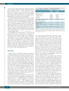
toxic effects were skin desquamation, headache and dizzi- ness, liver injury, edema, amenorrhea, hypertension and gastrointestinal disorders in the follow-up. Among patients who achieved an overall response, the median time to the treatment response was 34.87 days and the peak platelet count was 105.72×10⁹/L in the MSC-ITP-C+ group compared to 39.00 days and a peak platelet count of 80.62×10⁹/L in the MSC-ITP-C- group (Table 1).
To further clarify the mechanisms of the effect of ATRA in ITP, bone marrow MSC were isolated from ITP patients who had received 12 weeks of treatment. Compared with the baseline levels before ATRA therapy, the proliferative capacity, apoptosis, secretion of IL-1β and CXCL12, acti- vation of IL-1R/MyD88/NF-κB, ERK1/2 and p38 MAPK signaling pathways were altered, with trends similar to those in the in vitro studies in the patients from the MSC- ITP-C+ group after treatment (Figure 6D-H). In contrast, no significant differences were found between pre-treatment and post-treatment levels in patients from the MSC-ITP- C- group (Figure 6D-H).
Unsurprisingly, patients from the MSC-ITP-C+ group after treatment had overall increases in CXCL12 and megakaryocytes in the central marrow and decreases in the bone-associated marrow (Figure 6I, J), implying a CXCL12 gradient toward the sinusoids again. No signifi- cant differences of marrow CXCL12 distribution and spa- tial occupation of megakaryocytes were exhibited in the patients from the MSC-ITP-C- group after treatment (Figure 6I, J). The numbers of megakaryocytes and the density of vessels did not differ before and after treatment (Figure 6J). These data support the hypothesis that ATRA therapy contributed to changes in the location of CXCL12 production and the altered megakaryocyte location in the bone marrow niche for platelet generation.
Discussion
Thrombopoiesis, a complex biological process initiated by pluripotent hematopoietic stem cells in the bone mar- row, involves interactions with many cell types that con- tribute to the bone marrow niche, including osteoblasts, perivascular cells, endothelial cells, MSC, and various mature immune cells. Abnormalities during any stage of thrombopoiesis can influence platelet production.35 In the current study, we describe MSC impairment in the bone marrow of patients with ITP resulting from complement- derived perturbations in the IL-1β/IL-1R/NF-κB, ERK1/2 and p38 MAPK signaling pathways. Importantly, ATRA was found to decrease IL-1β mRNA expression and increase promoter DNA methylation. ATRA promoted functional recovery of MSC-ITP-C+ in vitro and in vivo, which facilitated the re-location of CXCL12 toward the vascular niche, enhanced megakaryocyte localization in the thrombopoietic niche and consequently promoted cir- culating platelet production (Figure 7).
Platelet destruction in ITP occurs through a variety of dif- ferent immune-mediated mechanisms.36 Current theories propose that antibodies against platelet-induced Fc-medi- ated phagocytosis, anti-GPIb/IX-mediated desialylation and the activation of cytotoxic lymphocytes are involved in the pathogenesis of ITP.1,37-39 The complement system has long been suspected to participate in platelet elimina- tion in patients with ITP.1-4 Activation of the complement cascade was found to play a role in the pathogenesis of ITP
Table 1. Responses and outcomes of immune thrombocytopenia patients with or without complement deposition on mesenchymal stem cells.
MSC-ITP-C+ MSC-ITP-C- (n=26) (n=32)
P value
0.013
0.266
0.363 0.190 0.048
Treatment response (%) Overall response
Complete response
Response No resonse
Blood component transfusion (%)
Significant bleeding *(%)
For patients with a response/ complete response
Time to response, days, median (range)
Peak platelet count, ×109/L, median
(range)
19 (73.08) 4 (15.38) 15 (57.69) 7 (26.92)
16 (61.54)
0 (0) 34.89 (23-52) 105.72 (62-201)
13 (40.63) 4 (12.50) 9 (28.13) 19 (59.37)
15 (46.88)
1 (3.13) 39.00 (27-53) 80.62 (37-112)
MSC-ITP-C+: mesenchymal stem cells with complement deposition from patients with immune
-
thrombocytopenia; MSC-ITP-C : mesenchymal stem cells without complement deposition from
patients with immune thrombocytopenia. *Significant bleeding was defined as bleeding events with a bleeding score of 3-4..
via complement-mediated generation of platelet micropar- ticles in the peripheral blood. Activation of the comple- ment system in the bone marrow of ITP patients was explored for the first time in this study. The observed enhanced deposition of C4d on MSC from patients with ITP was consistent with activation of the classical comple- ment pathway. The lack of observed increases in C3b dep- osition on MSC from ITP patients indicated that the alter- native complement pathway was not activated in bone marrow or that the extent of activation was at least less than the test threshold. Moreover, of 26 patients in the MSC-ITP-C+ group, five (19.2%) did not have detectable platelet antibodies, whereas of 32 patients in the MSC-ITP- C- group, 14 (43.7%) tested positive. Based on our results, there was not a one-to-one correspondence between the presence of autoantibodies and complement activation in the niche (Online Supplementary Table S3).
MSC residing in the bone marrow have long been believed to participate in regulating the balance between hematopoietic stem cell self-renewal and differentiation.40 In addition to their self-renewal properties and potential to differentiate, MSC play crucial roles in immune modulato- ry functions.41 We and others recently reported that MSC from ITP patients lose their conventional proliferation capacity resulting in defective immunoregulation.17-19,42,43 However, the mechanism underlying these abnormalities remains unknown. In the present study, we not only inves- tigated the enhanced complement deposition on the sur- face of MSC from ITP patients, but also explored the underlying genetic and molecular changes that give rise to deficiencies in MSC, which were associated with activa- tion of the complement cascade in bone marrow.
Complement components can enhance pro-inflamma- tory Toll-like receptor-mediated signaling in phagocytes, leading to increased production of IL-1β.20-22 IL-1β is criti- cally involved in several inflammatory diseases and its lev- els are elevated in many conditions characterized by com- plement overactivation. Furthermore, Martino et al. reported that IL-1β/IL-1R1/MyD88 signaling impairs MSC proliferation, migration and differentiation.44 However, it is not known whether and how complement and IL-1β are linked and their roles in MSC remain elusive. The biology
1672
haematologica | 2019; 104(8)
