
Page 163 - 2019_08-Haematologica-web
P. 163
Platelet GPVI and CLEC-2 in skin wound healing
attenuated by fibrinogen, and strongly inhibited by cross- linked fibrin compared to the migration through collagen (Online Supplementary Figure S7). Fibrinogen and fibrin showed a similar degree of inhibition for the migration of neutrophils from DKO mice (Online Supplementary Figure S7).
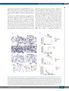
There was no significant difference in wound mono- cytes (Ly6C+) at day 1 post injury between DKO mice and WT (Online Supplementary Figure S4C and D). At day 3 post injury, DKO mice showed a significantly higher number of wound monocytes compared to all other strains (Figure 6A and B), which was reflected by a 4-fold increase in wound monocytes relative to day 1 post injury (Figure 6C). At day 9 post injury, monocytes within the wound remained at a low level in WT and single-knock- out mice (Figure 6D and E). Wound monocytes in DKO mice were decreased at this time compared to day 3 post injury (Figure 6C), but remained higher than other groups
(Figure 6D and E). In blood, Clec1bfl/flPf4-cre exhibited an increase in baseline circulating monocytes compared to WT and DKO mice (Online Supplementary Figure S4G, left). Blood monocytes were greatly reduced at day 3 and day 9 post injury in all groups compared to their unchal- lenged controls (Figure 6F). However, the level of blood monocytes in DKO mice was significantly higher than Clec1bfl/flPf4-cre mice at day 3 post injury (Online Supplementary Figure S4G, middle) and then all groups at day 9 post injury (Online Supplementary Figure S4G, right).
The influx of macrophages (F4/80+) in DKO mice was reduced at day 1 post injury compared to WT (Online Supplementary Figure S4E and F). At day 3 post injury, wound macrophages in Clec1bfl/flPf4-cre mice were elevat- ed, compared to all other strains (Figure 7A and B). A 6- fold increase in wound macrophages was observed in WT mice at day 3 relative to day 1 post injury (Figure 7C). DKO mice showed a significant reduction in
AB
D
C
E
F
Figure 6. A higher number of wound monocytes is observed during the inflammatory phase of repair in mice that lack both GPVI and CLEC-2. (A) Detection of monocytes (Ly6C+ cells; brown) in wound at day 3 post-injury. (B) Quantification of Ly6C+ cells in wound at day 3 post-injury (n=5-7). **P<0.01. (C) Comparison of Ly6C+ cells between day 1, day 3, and day 9 post-injury in WT and DKO mice. The symbols * and § indicate P<0.05 in WT and DKO mice, compared to the data at day 1 post injury, respectively. The bracket shows P<0.05 for the comparison between day 3 and day 9 post injury in §DKO mice. (D) Detection of Ly6C+ cells (brown) in wound at day 9 post injury. (E) Quantification of Ly6C+ cells in wound at day 9 post injury (n=6). *P<0.05; **P<0.01. (F) Comparison of blood monocyte counts between baseline, day 3, and day 9 post injury in each mouse strain. The symbols *, +, #, and § indicate P<0.05 in WT, Clec1bfl/flPf4-cre, Gp6-/-, and DKO mice, com- pared to their control, respectively. Sample numbers in unchallenged control, n=10; day 1, n=5; day 3, n=6-9; day 9 post injury, n=10-13, respectively. Graphs are presented as mean±Standard Error of Mean and analyzed by one-way ANOVA with Bonferroni’s multiple comparison test. Scale bar = 20 μm.
haematologica | 2019; 104(8)
1655
