
Page 162 - 2019_08-Haematologica-web
P. 162
S. Wichaiyo et al.
wound healing and is then cleared from the healing wound.
Platelet immunoreceptor tyrosine-based activation motif receptor deficiency reduces wound neutrophils and M1 macrophages during the inflammatory phase
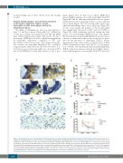
The influx of inflammatory cells was investigated at days 1, 3, and 9 post injury. Neutrophil (Gr-1+) infiltration on day 1 post injury was similar between WT and DKO mice (Online Supplementary Figure S4A and B). At day 3 post injury, DKO mice showed a significant impairment in neutrophil infiltration compared to WT and Clec1bfl/flPf4-cre mice but not Gp6-/- mice (Figure 5A and B). The decrease in neutrophil infiltration was confirmed using anti-Ly6G antibody clone 1A8 (data not shown). A 2- fold increase in wound neutrophils was observed in WT but not in DKO mice at this time relative to day 1 post
injury (Figure 5C). At day 9 post injury, DKO mice showed higher numbers of wound neutrophils than WT (Figure 5D and E), although neutrophil level was signifi- cantly decreased in both groups relative to day 1 and day 3 post injury (Figure 5C). Blood neutrophil counts were similar in unchallenged mice across all groups (Figure 5F). At day 3 post injury, the number of blood neutrophils was significantly decreased in WT and Clec1bfl/flPf4-cre (Figure 5F) while remaining unaltered during the time course of wound healing in DKO and Gp6-/- mice (Figure 5F). The decrease in neutrophil infiltration was not due to a defect of chemoattractants at the wound site as meas- ured by the presence of chemokine CXCL-1 (Online Supplementary Figure S6A and B) or platelet factor 4 (PF4) (Online Supplementary Figure S6C and D). In vitro migra- tion towards N-formyl-methionyl-leucyl-phenylalanine (fMLP) using bone marrow-derived neutrophils demon- strated that migration of neutrophils from WT mice was
A
B
C
DE
F
Figure 5. Neutrophil influx is decreased during the inflammatory phase of wound healing following platelet CLEC-2 and GPVI double-deletion. (A) Detection of neu- trophils (Gr-1 staining; brown) in wound at day 3 post injury. (B) Quantification of neutrophils (Gr-1+ cells) in wound at day 3 post injury. *P<0.05; **P<0.01. (C) Comparison of Gr1+ cells between day 1, day 3, and day 9 post injury in wild-type (WT) and DKO mice. P<0.05 in *WT and §DKO mice, compared to the data at day 1 post injury, respectively. The bracket shows P<0.05 for the comparison between day 3 and day 9 post injury in *WT and §DKO mice, respectively. (D) Detection of neutrophils (Gr-1 staining; brown) in wound at day 9 post injury. (E) Quantification of neutrophils (Gr-1+ cells) in wound at day 9 post injury. *P<0.05. (F) Comparison of blood neutrophil counts between baseline, day 3, and day 9 post injury in each mouse strain. P<0.05 in *WT and +Clec1bfl/flPf4-cre mice, compared to their con- trol, respectively. Sample numbers in unchallenged control = 10, day 1 = 5, day 3 = 6-9, and day 9 post injury = 9-13, respectively. Graphs are presented as mean±Standard Error of Mean and analyzed by one-way ANOVA with Bonferroni’s multiple comparison test. Scale bar = 20 μm.
1654
haematologica | 2019; 104(8)
