
Page 160 - 2019_08-Haematologica-web
P. 160
S. Wichaiyo et al.
blood cells and plasma proteins into the wound21 where they come in contact with TF,5,22 which activates the extrinsic pathway of blood coagulation. An increase in fibrinogen deposition was particularly marked in the granulation tissue at day 3 post injury (Figure 4B and C) with no significant alteration in TF expression in DKO mice (Online Supplementary Figure S3A and B). Fibrin con-
tent in the wound scab was similar between WT and DKO mice at day 1 post injury (Online Supplementary Figure S3C and D), but was significantly increased in the DKO mice at day 3 post injury, compared to WT and Clec1bfl/flPf4-cre mice but not to Gp6-/- mice, which exhibit- ed a more moderate vascular leakage (Figure 4D and E). At day 9 post injury, fibrin was mainly located on the
AB
E
C
D
F
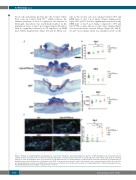
Figure 3. Enhanced re-epithelialization and angiogenesis occur at the early phase of wound healing in the absence of GPVI and CLEC-2. (A) Hematoxylin & Eosin staining at day 3 post-injury (n=6-9). Dotted line indicates hyperplastic coverages. Black arrow points to wound edge. Red arrow indicates gap between epithelial tongues. S: scab; G: granulation tissue. Scale bar=500 μm. (B) Measurement of re-epithelialization. (C) Measurement of wound contraction. (D) Quantification of granulation tissue area. (E) Detection of endothelial cells (CD31+ cells; green) in wound area at day 3 post injury. Hoechst counterstains nuclei (blue). Scale bar=50 μm. (F) Quantification of CD31+ area within the wound at day 3 post injury (n=5-6). Graphs are presented as mean±Standard Error of Mean and analyzed by one- way ANOVA with Bonferroni’s multiple comparison test. *P<0.05.
1652
haematologica | 2019; 104(8)
