
Page 159 - 2019_08-Haematologica-web
P. 159
Platelet GPVI and CLEC-2 in skin wound healing
These results demonstrate that deletion of both CLEC- 2 and GPVI accelerates skin wound closure, enhances re- epithelialization and angiogenesis, and reduces scar for- mation.
GPVI and CLEC-2 maintain vascular integrity during the inflammatory phase of wound repair
The redness surrounding the wound in DKO mice in the inflammatory phase is indicative of increased vascular leakiness. Macroscopic examination of the skin at day 3 post injury demonstrated vasodilation and bleeding into the wound as well as into the surrounding skin in DKO mice, with less severe vascular leakage seen in Gp6-/- mice (Figure 4A). Hematoxylin & Eosin (H&E) staining con- firmed the extravasation of red blood cells (RBC) in the dermis at the edge of the wound at day 3 post injury in
both DKO and Gp6-/- mice (Online Supplementary Figure S2A). Clearance of extravascular RBC was observed at day 9 post injury in all groups (Online Supplementary Figure S2B). Due to blood/lymphatic mixing phenotype in DKO, and to a lesser degree in Clec1bfl/flPf4-cre mice, RBC were also present in lymphatic vessels (podoplanin+) (Online Supplementary Figure S2C).
These observations demonstrate marked impairment of vascular integrity during the inflammatory phase of wound repair in DKO mice, with a milder phenotype in Gp6-/- mice. Vascular leakage is diminished in later phases when inflammation subsides.
GPVI and CLEC-2 deficiency increases fibrin(ogen) deposition during the inflammatory phase of repair
Increased vascular permeability results in leakage of
AB
C
DE
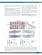
Figure 2. Deletion of platelet immunoreceptor tyrosine-based activation motif (ITAM) receptors accelerates skin wound repair process. Mice were subjected to a full-thickness excisional skin wound and wound closure was monitored for nine days after injury. (A) Macroscopic appearance of wound at indicated time points. (B) Changes of wound size over nine days post injury (n=10-13). (C) Hematoxylin & Eosin staining at day 9 post-injury (n=9-13). a: length of hyperplastic epidermis; b: inter-subcutaneous distance. Scale bar=200 μm. (D) Measurement of the length of hyperplastic epidermis. (E) Measurement of inter-subcutaneous distance. All graphs are presented as mean±Standard Error of Mean (SEM). Kinetics of wound closure (B) are analyzed by two-way ANOVA with Bonferroni’s multiple comparison test. *P<0.05; **P<0.01. *WT versus DKO. +Clec1bfl/flPf4-cre versus DKO. #Gp6-/- versus DKO. Other parameters are analyzed by one-way ANOVA with Bonferroni’s multiple comparison test. *P<0.05.
haematologica | 2019; 104(8)
1651
