
Page 158 - 2019_08-Haematologica-web
P. 158
S. Wichaiyo et al.
However, the morphometric analysis of skin histology at this time point revealed a significantly smaller scar in DKO mice, as shown by a shorter length of hyperplastic epidermis compared to WT and Clec1bfl/flPf4-cre mice (Figure 2C and D) and a narrower inter-subcutaneous gap than WT mice (Figure 2C and E).
At day 3 post injury, re-epithelialization was observed in all groups (Figure 3A). However, this process was enhanced in DKO mice compared to WT and Clec1bfl/flPf4- cre mice, but not Gp6-/- mice (Figure 3B). There was no sig-
nificant difference in wound contraction between all test- ed groups (Figure 3C). DKO mice also had a larger area of granulation tissue compared to WT mice (Figure 3A and D). Improved wound healing was associated with enhanced angiogenesis as assessed by the increase in CD31+ area in DKO animals at day 3 post injury (Figure 3E and F), although the density of blood vessels (CD31+) and lymphatic vessels (podoplanin+) in unchallenged skin was similar among all groups (Online Supplementary Figure S1E and F).
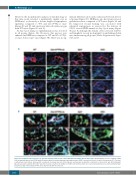
A
B
Figure 1. Podoplanin-expressing cells are present at perivascular area in contact with platelets during skin wound repair. (A) Immunofluorescence staining of NG2 (red), podoplanin (green) and CD41 (white) illustrates platelets and podoplanin-expressing pericytes (NG2+) around blood vessel at day 3 after injury (n=5-7). Hoechst counterstains nuclei (blue). Arrow points to platelets at perivascular site. Star indicates extravascular localization of platelets. Scale bar=20 μm. (B) Podoplanin (green) was double-stained with either vimentin (red; top) or Ly6C (red; middle) or F4/80 (red; bottom), which are located around blood vessel (surrounded by NG2+ pericytes) at day 3 after injury (n=4-5). Scale bar=20 μm. BV: blood vessel.
1650
haematologica | 2019; 104(8)
