
Page 168 - 2019_06-Haematologica-web
P. 168
A.C. Glembotsky et al.
in platelets from a third pedigree harboring the T219Rfs*8 mutation (pedigree D) (Figure 3A). Western blot analysis of platelet samples confirmed a marked decrease in TLT-1 content (Figure 3B), revealing for the first time downregu- lation of a member of the family of ITIM-bearing recep- tors in patients with RUNX1 mutations. We have previ- ously shown that patients from this pedigree display par- tial deficiency of α-granules,8 where TLT-1 is located, and this finding was associated with a mild decrease in α-gran- ule protein TSP-1.8 Heterogeneous platelet content of other α-granule proteins was demonstrated by other authors, including reduced levels of PF4, which is regulat- ed by RUNX1,24 and preserved levels of b-TG and PDGF.25 More recently, RUNX1 deficiency has been linked to abnormal ER-to-Golgi trafficking and protein sorting lead- ing to low von Willebrand factor (vWF) α-granule content.26 Although down-regulated TREML1 gene expression secondary to RUNX1 loss-of-function seems to be the major mechanism leading to the low TLT-1 found in this study, we cannot exclude the possibility that α- granule defects could contribute in part to the profound reduction in TLT-1 protein.
Whereas incubation of normal platelets with a blocking anti-TLT-1 antibody inhibited thrombin-induced platelet aggregation, as previously reported,19 it had no effect on thrombin-induced aggregation of FPD/AML platelets (Figure 3C), further demonstrating the absence of relevant amounts of TLT-1 in patient platelets. Conversely, consis- tent with the functional role of sTLT-1 in platelet activa- tion,17 and as previously shown for platelet adhesion and actin polymerization on fibrinogen matrices,27 a recombi- nant soluble fragment (rsTLT-1) was able to potentiate thrombin-induced fibrinogen binding to normal platelets (Figure 3D). In the absence of rsTLT-1, thrombin-induced fibrinogen binding was lower in patients from pedigree D compared to controls, as previously reported.8 This defect was partially corrected by incubation of patient platelets with rsTLT-1 (Figure 3D), pointing to a role for TLT-1 defi- ciency in the platelet function defect. However, several other abnormalities are also involved, possibly contribut- ing to the variability in platelet aggregation tests among patients (Table 1).
As shown in this work, TREML1 gene expression levels are low in normal early megakaryopoiesis and increase markedly along MK maturation reaching higher expression levels in mature MK. Double immunofluorescence labeling of TLT-1 and vWF in mature (CD41+CD42+) MK from one
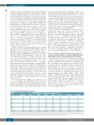
Table 2. Surface platelet glycoprotein expression.
Relative fluorescence intensity (patient/control ratio)
control and 2 patients (DII-1 and DIII-3) confirmed that TLT-1 is mainly localized in α-granules (Figure 4), as previ- ously reported.14 However, part of TLT-1 does not co-local- ize with vWF and is present in a different subpopulation of α-granules. Interestingly, a distinct staining pattern of TLT- 1 and P-selectin was recently shown in mouse and human platelets and mouse MK, suggesting differential compart- mentalization of these proteins within α-granules,15 as pre- viously shown for other proteins packaged in platelets.28 TLT-1-positive granules were less abundant or absent in patient compared to control MK (Figure 4).
The role of TLT-1 in normal MK is not known. TLT-1- deficient mice display a 20% decrease in platelet counts,17 although the underlying mechanism has not been explored. In order to gain insight into this, we incubated human cord blood or leukapheresis-derived MK with a blocking anti-TLT-1 antibody or a control IgG. There was no significant difference in megakaryocyte output and maturation in the presence of anti-TLT-1 compared to control, whereas proplatelet formation was reduced (Online Supplementary Figure S3), suggesting TLT-1 may have a role in normal proplatelet formation. However, fur- ther study will be required to definitively establish this issue and to determine whether TLT-1 deficiency con- tributes to defective platelet production in FPD/AML.
Levels of integrin subunit α2 and collagen adhesion are decreased in platelets in familial platelet disorder with predisposition to acute myelogenous leukemia
Patient
GPIIa (b1) AII-1 0.48
AII-2 0.65 BII-2 nd BIII-1 nd DII-1 0.43 DIII-1 0.77 DIII-3 0.65
Ref. range 0.81-1.19
GPVI GPIIb
nd 0.98
nd 0.96 nd 0.90 nd 0.94
0.79 0.79 0.89 0.79 0.80 0.98 0.73-1.27 0.78-1.22
GPIIIa GPIb
1.09 1.44
1.07 1.87
1.01 1.99
0.97 2.37
1.02 1.51
0.91 1.42
1.03 1.58
0.78-1.22 0.65-1.35
GPIX α5 integrin 1.22 nd
1.47 nd 1.85 nd 2.07 nd 1.84 0.95 1.74 1.29 1.28 0.98
0.81-1.19 0.85-1.12
α6 integrin nd
nd
nd
nd
0.78
1.32
0.78
0.70-1.55
Transcript levels of ITGA2, coding for the α2 integrin subunit of collagen receptor α2b1, were also shown to be decreased in platelets from pedigree D by qPCR (Figure 5A). Accordingly, surface expression of α2 (GPIa) was sub- stantially reduced, as revealed by analysis of platelet-rich plasma (Figure 5B) and whole blood flow cytometry (Online Supplementary Table S5), and further confirmed by western blot (Figure 5C). The reduction in surface α2 was associated with a decrease in platelet surface expression of the heterodimeric b1 subunit (GPIIa), whereas GPVI, GPIIbIIIa, GPIb-IX were preserved (Table 2), indicating a selective abnormality in the α2b1 complex. The reduction in b1 is probably due to the concomitant decrease in its α2 partner, as b1 subunit (ITGB1) mRNA levels were pre- served (Online Supplementary Figure S4). In addition, platelet surface levels of α5 and α6 integrin subunits, which also heterodimerize with b1, were normal or in the lower normal limit (Table 2).
GP: glycoprotein. Ref. ; reference; nd; not done. GPIIa represents the b1 integrin subunit of the collagen receptor α2b1. Relative fluorescence intensity (RFI) was calculated as the ratio between the corresponding antibody and the isotypic control.Results are expressed as the ratio between RFI in each patient and a simultaneously assayed control sample. The reference range was established by the Mean ± two Standard Deviations of ten healthy subjects who were simultaneously studied.
1250
haematologica | 2019; 104(6)
