
Page 63 - 2019_04-Haematologica-web
P. 63
133±10 87±7 102±7 72±17 1 (9) 1 (9)
HbAA (n=9, 82%) HbAS (n=2, 18%) 13.6±1.3 1.3±0.5 60±25 0.6±0.5 39.3±31.1 190±31
<0.01 <0.001 <0.001 0.51
0.26++ 0.85++
Impaired CVR in adults with sickle cell disease
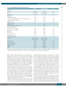
Table 1. Patient characteristics and baseline measurements. Characteristic
Age, years
Sex Bodyweight, kg Ethnicity, n (%)
South America: Suriname
Western Africa: Benin, Congo, Ghana, Guinea, Nigeria, Sierra Leone Caribbean: Dutch Antilles, Jamaica
Europe: Turkey
Medication & therapy
Hydroxyurea, n (%)
Chronic blood transfusion therapy, n (%) Cardiovascular risk factors
Systolic blood pressure, mmHg
Diastolic blood pressure, mmHg
MAP, mmHg
Heart rate, bpm
Nicotine smokers, n (%) Cannabis smokers, n (%) Hematologic characteristics Genotype
Hemoglobin, g/dL Reticulocyte count, % Reticulocyte count, #10 /L Bilirubin total, mg/dL 9 ASAT, U/L
LDH ++ 2
infarcts (SCI).3,4 Although SCI were once thought to be benign, SCI volume is associated with reduced cognitive performance in children with SCD,5 and a 14-fold increased risk of overt stroke in pediatric SCD.6 SCI risk increases relentlessly with age,7,8 reaching a prevalence of 50% by the age of 30.9 There is currently no treatment for SCIs in adults, although efforts to reduce their incidence by blood transfusions have been made.10 Nevertheless, identifying modifiable risk factors and predictors of these lesions is a focus of current research in adult SCD.
Cerebrovascular reserve (CVR) is a measure of the via- bility of cerebral vessels to respond to a vasoactive stimu- lus and is often used to study hemodynamic status in neu- rovascular diseases.11 CVR is defined as the remaining vasodilating capacity of the cerebral arterioles in response to an exogenous stimulus such as CO2 or acetazolamide.12 Recent studies suggest that impaired CVR predicts loca- tions of lesions at one year follow up13 and the risk of stroke in steno-occlusive disease,14,15 providing compelling evidence that this hemodynamic marker can inform future cerebral damage observed on MRI.
However, the predictive value of CVR in patients with SCD has not yet been shown. Previous cross-sectional studies have observed reduced CVR in children and adults
Controls
n=11
37.4 ± 15.4
6 men, 5 women 76 (14)
7 (64) 2 (18) 1 (9) 1 (9)
Patients with SCD
n=36
31.9 ± 11.3
23 men, 12 women 70 (18)
19 (54) 11 (31) 4 (11) 1 (3)
13 (37)
3 (9)
121±10 71±8 87±7 76±11 9 (26) 4 (11)
P* 0.52
0.46++ <0.01
0.59 0.39 0.83 0.38
-
-
-
-
HbSS (n=31, 89%) HbS 0 (n=4, 11%) 8.8±1.4↓
-
8.9±4.2↑ 261±108↑ 3.1±2.0↑ 48.1±16.2↑ 459±165↑
-
<0.001 <0.001 <0.001 <0.001 <0.05 <0.05
with SCD without a history of overt stroke,16–20 and we have recently shown dilated cerebral vessels at baseline in SCD.21 Together, these findings suggest that high resting blood flow demands are met by vasodilation. Vasodilation at rest will limit further dilation in times of increased demand, which poses a risk for ischemia. Examples of such risk in SCD include infection, fever, acute anemic events,22 and obstructive sleep apnea.23 CVR was previous- ly associated with anemia,20 and dilatory function of the vessels could additionally be impaired in SCD due to vas- cular inflammation, low nitric oxide and abnormal endothelial function. Since the brain cannot store its own oxygen, it must maintain constant perfusion, and inade- quate oxygen delivery by increases in demand or low hemoglobin may cause ischemia and SCIs due to a lack of vasodilatory reserve. We hypothesized that CVR is lower in patients with SCD compared to healthy controls, and that SCIs are related to low CVR.
The objective of this study was to investigate regional CVR measurements in SCD and to investigate the associ- ation between CVR and the presence and volume of exist- ing ischemic lesions in SCD. In the current study, we used arterial spin labelling (ASL), a non-contrast perfusion MRI method, to assess whole brain CBF prior to and following
c test. *T-test, or Wilcoxon signed rank test was used to test the statistical significance of the difference as appropriate. MAP: mean arterial pressure: [(2 *DiastolicBP) + SystolicBP)]/ 3 mmHg. ↑ above healthy reference / ↓ below healthy reference. LDH: Lactate dehydrogenase.
haematologica | 2019; 104(4)
691
