
Page 58 - 2019_04-Haematologica-web
P. 58
C.J. Mercadante et al.
AB
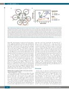
Figure 6. Mathematical modeling predicts increased excretion rates in treated Trfhpx/hpx mice. (A) The iron (Fe) homeostasis scheme used for mathematical modeling of body Fe levels from Figure 3B. Closed polygons represent liver, spleen, red blood cells (RBC), bone marrow (BM), duodenum (Duo), or ‘rest of body’ (Rest), the last comprising stomach, intestines except the duodenum, integument, muscles, heart, fat, lungs, kidneys, brain, and reproductive organs. The circle represents plasma. Dietary Fe is absorbed into the duodenum (solid dark green arrow) and exported to plasma by ferroportin (solid black arrow). Plasma Fe, in states of Fe homeostasis, exists as Fe-loaded transferrin (FeTF) which is imported into all compartments (solid light green arrows) except RBC or, in states of Fe excess, as non-transferrin- bound iron (NTBI) which is imported into the liver and rest of the body (solid blue arrows). Bone marrow Fe is incorporated into RBC and recycled in the spleen (solid red arrows); direct Fe transfer from the bone marrow to the spleen represents recycling of immature erythroid cells in the spleen (solid red arrows). Fe can be exported from compartments by ferroportin (solid black arrows). FeTF stimulates hepcidin expression (dashed black arrow), which inhibits ferroportin-dependent Fe export from compartments (dashed blunt-ended lines). Erythropoietin (EPO) stimulates FeTF import into the bone marrow, transfer of Fe from the bone marrow to RBC, and suppression of hepcidin activity. EPO is suppressed by increased RBC Fe levels. Fe can be excreted from the liver, duodenum, and the rest of the body (solid magenta lines). (B) Results of mathematical modeling. Experimental values from Figure 3B are shown as circles. Modeled values are shown as lines.
(Figure 5F). Measurement of total fecal iron levels indicat- ed no significant difference between any experimental group (Online Supplementary Figure S4). This was expected given that total fecal iron levels are affected by multiple factors beyond excretion, including dietary iron levels and iron absorption. Analysis of 59Fe-labeled compounds in feces was not possible - 59Fe levels were low at the time of collecting the feces and decayed significantly once all mice had been processed in the excretion study. As fecal ferritin levels have been reported to reflect body iron lev- els19, we next measured fecal ferritin levels by ELISA. Similar to our estimates of fecally excreted iron (Figure 5F), fecal ferritin levels were increased in all Trfhpx/hpx mice relative to Trf+/+ mice and increased in untreated Trfhpx/hpx mice relative to treated Trfhpx/hpx mice (Figure 5G, Online Supplementary Figure S5). We next attempted to assess fer- ritin iron levels in fecal samples. Using mouse liver lysates, we established that the same iron stain used for histology could be used to detect a species very abundant in Trfhpx/hpx mouse liver that comigrated with ferritin heavy and light chain under native PAGE conditions (Online Supplementary Figure S6). However, native PAGE of fecal lysates failed to reveal stainable iron (data not shown).
Mathematical modeling predicts increased excretion rates in treated Trfhpx/hpx mice
We also analyzed body iron levels from Figure 3B using our mathematical model of iron homeostasis, summa- rized in Figure 6A.20 The goal here was to explore the decrease in body iron content in transferrin-treated Trfhpx/hpx mice from 4 to 6 months (Figure 3B,C) - our excre- tion study was halted at 4.5 months as not all untreated Trfhpx/hpx mice survived to 6 months. Using the model, we could fit body iron levels from Trf+/+ and untreated Trfhpx/hpx mice with good agreement between experimental and modeled values (Figure 6B). However, we were unable to
reproduce iron levels in 6-month old untreated Trfhpx/hpx mice. As most untreated Trfhpx/hpx mice die before 6 months, iron loss at this age may not reflect physiological excretion but rather cell death secondary to severe iron excess or other long-term adverse effects of transferrin deficiency. When we simulated body iron levels in trans- ferrin-treated Trfhpx/hpx mice, we could reproduce body iron levels up to 4 months but were initially unable to repro- duce the decrease in body iron levels from 4 to 6 months. We considered that the decrease in iron levels from 4 to 6 months in transferrin-treated Trfhpx/hpx mice required an increase in excretion from one or more compartments or a decrease in absorption starting at 4 months. The small- est change in absorption or excretion that fitted the data was a four-fold increase in excretion from the duodenum and ‘rest of body’. Given that ‘rest of body’ in our model comprised other gastrointestinal regions including jejunum, ileum, and large intestine, this supports our 59Fe- based studies indicating that the gastrointestinal tract is the main route of excretion.
Discussion
In this study, we exploited our initial observation that transferrin treatment decreases the concentrations of iron in the organs of Trfhpx/hpx mice to explore the basis of iron excretion. For this objective, the Trfhpx/hpx model has some key advantages over other mouse models of common human diseases of iron excess such as hereditary hemochromatosis and β-thalassemia. First, Trfhpx/hpx mice develop more severe iron excess than most other models. We anticipated that the severity of iron excess would sig- nificantly increase iron levels in potential routes of excre- tion such as feces and urine. Second, the primary defect in Trfhpx/hpx mice can be rapidly corrected pharmacological-
686
haematologica | 2019; 104(4)
