
Page 60 - 2019_04-Haematologica-web
P. 60
C.J. Mercadante et al.
untreated Trfhpx/hpx mice relative to those in Trf+/+ mice at 2.5 months of age (Online Supplementary Figure S7), the rate at which biliary iron is eliminated from the body is not only influenced by biliary iron levels. Dietary iron deficiency can alter rates of bile synthesis in rats.53 In rats, iron can also undergo enterohepatic circulation, the process by which substances excreted in bile are reabsorbed by the small intestine and transported back to the liver.54 A study of the role of hepatobiliary iron excretion would require measurement of bile synthesis rates and iron levels and rates of enterohepatic circulation in multiple animal mod- els of iron excess and deficiency. Analysis of the biochem- ical form of biliary iron is also warranted, as this may indicate a potential mechanism for iron excretion. Iancu et
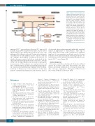
Figure 7. Model of iron absorption and excretion. Dietary non-heme iron (Fe) is absorbed by enterocytes in the small intestine. Enterocytes export Fe into the blood; this process can be inhibited by hepcidin. Fe is then transported to the liver for storage, excretion, or distribu- tion to other organs in the body. Fe can be excreted by the liver into bile and transported into the small intestine, where it can undergo enterohepatic cir- culation or can be eliminated from the body via the feces. Fe can also be excreted from the body by turnover of epithelial cells lining the intestines or from minor trauma to intestinal epitheli- um leading to blood loss. Dashed lines indicate minor routes of Fe excretion, which include blood loss, exfoliation of dead skin, and excretion via the urine. For the sake of simplicity, not all organs or pathways of Fe transport are shown, including those that mediate heme Fe absorption.
al. observed electron-dense material within bile canaliculi similar in appearance to hemosiderin.55 This type of study, along with a study of the contribution of epithelial turnover to iron excretion, would also need to be per- formed in both male and female subjects given that 59Fe excretion rates were decreased in untreated male but not female Trfhpx/hpx mice (Figure 4E).
Acknowledgments
The authors would like to thank Joseph Orchardo and David Murray for assistance with metal measurements and Iqbal Hamza and Anatoly Zhitkovich for reviewing the manuscript. This work was supported by NIH grants DK84122 and DK110049 (TBB) and GM080219 (PM).
References
1. Coffey R, Ganz T. Iron homeostasis: an anthropocentric perspective. J Biol Chem. 2017;292(31):12727–12734.
2. Dev S, Babitt JL. Overview of iron metabo- lism in health and disease. Hemodial Int Int Symp Home Hemodial. 2017;21 (Suppl 1)S6–S20.
3. Muckenthaler MU, Rivella S, Hentze MW, Galy B. A red carpet for iron metabolism. Cell. 2017;168(3):344–361.
4. Papanikolaou G, Pantopoulos K. Systemic iron homeostasis and erythropoiesis. IUBMB Life. 2017;69(6):399–413.
5. Wallace DF. The regulation of iron absorp- tion and homeostasis. Clin Biochem Rev. 2016;37(2):51–62.
6. Brissot P, Cavey T, Ropert M, Guggenbuhl P, Loréal O. Genetic hemochromatosis: pathophysiology, diagnostic and therapeu- tic management. Presse Med. 2017;46(12 Pt 2):e288–e295.
7. Kawabata H. The mechanisms of systemic iron homeostasis and etiology, diagnosis, and treatment of hereditary hemochro- matosis. Int J Hematol. 2018;107(1):31–43.
8. Asadov C, Alimirzoeva Z, Mammadova T,
Aliyeva G, Gafarova S, Mammadov J. β- thalassemia intermedia: a comprehensive overview and novel approaches. Int J Hematol. 2018;108(1):5-21.
9. Cappellini MD, Motta I. New therapeutic targets in transfusion-dependent and -inde- pendent thalassemia. Hematol Am Soc Hematol Educ Program. 2017;2017(1):278– 283.
10. Gupta R, Musallam KM, Taher AT, Rivella S. Ineffective erythropoiesis: anemia and iron overload. Hematol Oncol Clin North Am. 2018;32(2):213–221.
11. Bu JT, Bartnikas TB. The use of hypotrans- ferrinemic mice in studies of iron biology. Biometals. 2015;28(3):473–480.
12. Bartnikas TB, Andrews NC, Fleming MD. Transferrin is a major determinant of hep- cidin expression in hypotransferrinemic mice. Blood. 2011;117(2):630–637.
13. Raja KB, Pountney DJ, Simpson RJ, Peters TJ. Importance of anemia and transferrin levels in the regulation of intestinal iron absorption in hypotransferrinemic mice. Blood. 1999;94(9):3185–3192.
14. Buys SS, Martin CB, Eldridge M, Kushner JP, Kaplan J. Iron absorption in hypotrans- ferrinemic mice. Blood. 1991;78(12):3288– 3290.
15. 16.
17. 18.
19.
20.
21.
Parmar JH, Mendes P. A computational model to understand mouse iron physiolo- gy and diseases. bioRxiv. 2018;323899. Kautz L, Jung G, Valore EV, Rivella S, Nemeth E, Ganz T. Identification of ery- throferrone as an erythroid regulator of iron metabolism. Nat Genet. 2014;46(7):678–684.
Stevens AR, White PL, Hegsted DM, Finch CA. Iron excretion in the mouse. J Biol Chem. 1953;203(1):161–165.
Chappelle E, Gabrio BW, Stevens AR, Finch CA. Regulation of body iron content through excretion in the mouse. Am J Physiol. 1955;182(2):390–392.
Skikne BS, Whittaker P, Cooke A, Cook JD. Ferritin excretion and iron balance in humans. Br J Haematol. 1995;90(3):681– 687.
Parmar JH, Davis G, Shevchuk H, Mendes P. Modeling the dynamics of mouse iron body distribution: hepcidin is necessary but not sufficient. BMC Syst Biol. 2017;11(1): 57.
Casu C, Oikonomidou PR, Chen H, et al. Minihepcidin peptides as disease modifiers in mice affected by β-thalassemia and poly- cythemia vera. Blood. 2016;128(2):265– 276.
688
haematologica | 2019; 104(4)
