
Page 114 - 2019_04-Haematologica-web
P. 114
R. Polak et al.
MTAB-3466. Microarray data of BCP-ALL blasts are available in the Gene Expression Omnibus database.
Informed consent was provided according to the Declaration of Helsinki. Use of left-over materials for research purposes was approved by the Institutional Review Board (IRB) of the University Medical Center Rotterdam; IRB approval file num- ber MEC 2004-203.
Functional assays
For protein quantification, both western blot analysis and reverse phase protein arrays (RPPA) were used. Vps34 promoter activity was studied using a reporter construct consisting of a 1.4 kB region of the Vps34 promoter upstream of the Gaussia luciferase gene. Cell viability was quantified using MTT cyto- toxicity assays or flow cytometry-based Annexin V - Propidium Iodide assays. Silencing of genes was achieved using transfec-
tion of specific siRNAs or a lentiviral knockdown approach using the pLKO.1 Mission vector containing a puromycin selec- tion marker. Autophagy levels (number and volume of LC3B- positive vesicles) were quantified using confocal scanning microscopy (Leica SP5).
Statistical analysis
Statistical analysis of microarray data of paired CB-CD34+ cells was performed using a linear mixed model. Microarray data of primary ALL samples were analyzed using LIMMA. Functional analysis of differential gene expression was performed using QIAGEN’s Ingenuity Pathway Analysis. Both the Student t-test and the Student paired t-test were used when applicable. Bar graphs represent the mean of biological replicates. Error bars rep- resent standard error of mean (SEM). Further details of the meth- ods used are available in the Online Supplementary Appendix.
ABC
D
E
F
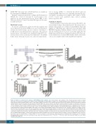
Figure 4. Vps34 is essential for the survival of ETV6-RUNX1-positive leukemic cells. (A) Schematic representation of the known transcript variants of Vps34 (Ensemble Genome Browser; ENSG00000078142) and the shVps34#1 and shVps34#2 recognition sites. (B) ETV6-RUNX1-positive (REH) BCP-ALL cells were lentivi- rally transduced with scrambled shRNA control, or two distinct shRNA constructs to silence Vps34 expression. Western blot analysis was performed with an antibody against Vps34 or β-actin to visualize the knockdown of Vps34 in REH cells. A representative experiment is shown in which increasing concentrations of virus were used to emphasize the specificity of Vps34 knockdown. (C) Data were quantified and are depicted as the relative Vps34 expression in comparison to the expression in cells transduced with scrambled (non-silencing) controls. (D) ETV6-RUNX1-positive (REH and REHS1) and ETV6-RUNX1-negative (NALM6) BCP-ALL cells were lentivirally transduced with scrambled shRNA control (NSC) or two distinct Vps34 shRNA constructs. NI: non-infected cells. Cells were cultured for 18 days. To deter- mine the effect on proliferation, cell counts were performed every 2-3 days. Representative graphs are shown (n=3). (E and F) ETV6-RUNX1-positive (REH) BCP-ALL cells were lentivirally transduced with scrambled shRNA control (NSC) or two distinct shRNA constructs to silence Vps34 expression. After seven days of culture, flow cytometrical analysis was performed to determine the effect of Vps34 knockdown on survival and cell-cycle progression. (E) The percentage of viable (AnnexinV-pos- itive, Propidium-Iodide negative), actively cycling cells was determined using DyeCycle. Data are shown as the percentage of cells in S, G2 M phase (n=2; *P≤0.05). Error bars represent Standard Error of Mean (SEM). (F) The percentages of early apoptotic (AnnexinV-positive, Propidium-Iodide negative) and late apoptotic (Propidium-Iodide positive) cells were determined seven days after transduction (n=2, **P≤0.01, ***P≤0.001). Error bars represent SEM. See also Online Supplementary Figure S7.
742
haematologica | 2019; 104(4)
