
Page 113 - 2019_04-Haematologica-web
P. 113
Autophagy drives ETV6-RUNX1-positive leukemia
network underlying the persistence and maintenance of ETV6-RUNX1 BCP-ALL remains to be elucidated.
In the present study, we address the role of autophagy in ETV6-RUNX1-driven leukemia. Autophagy is a cellular recycling system in which unwanted or damaged cellular components are degraded and recycled. The core autophagy-regulating complex includes Vps34, Beclin-1, and Vps15.21,22 Although autophagy can sustain cell sur- vival during stress conditions, it can also result in cell death because of progressive cellular consumption.23 Whether autophagy plays an initiating or suppressive role in cancer is a question of debate and most likely depends on the (onco)genetic context of cells.24,25 This potential dual role of autophagy in cancer highlights the importance of studies on the context-specific role and the functional importance of autophagy in neoplastic processes before the start of autophagy-based therapeutic interventions. We show here that ETV6-RUNX1 targets the autophagy process, which in turn affects sensitivity to L- Asparaginase, a key enzyme used in the treatment of ALL
that affects the asparagine (and to a lesser extent gluta- mine) levels in cells.
Methods
Transduction and gene expression profiling of primary cells
CD34-positive hematopoietic progenitor cells (CB-CD34+ cells) were derived from human cord blood and transduced with retro- virus expressing ETV6-RUNX1 and eGFP. DAPI-CD34+ GFP+ CB- CD34+ cells were sorted using a BD ARIA II sorter. After sorting, cells were lysed and RNA was extracted and subsequently linearly amplified.
Bone marrow aspirates were obtained from children with newly diagnosed BCP-ALL prior to treatment. Leukemic blasts were collected and processed as previously described.
Affymetrix GeneChip HG-U133-Plus-2.0 microarrays were used for all samples. Microarray data of CB-CD34+ cells are avail- able in the ArrayExpress database under accession number E
AB
CDE
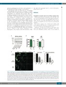
Figure 3. Autophagy levels are high in ETV6-RUNX1-positive B-cell precursor acute lymphoblastic leukemia (BCP-ALL) and regulated by ETV6-RUNX1 and Vps34.
(A) Western blot analysis was performed to determine the expression levels Vps34, p62 (sequestosome 1), and LC3B in ALL cell lines. PIK3R4 was used as a loading control. (B) Quantification of protein levels of Vps34 and p62 measured by reverse phase protein array (RPPA) in 30 ETV6-RUNX1-positive primary BCP-ALL patient samples (ETV6-RUNX1+), and 29 B-Other primary BCP-ALL patient samples (BO). Data are means±Standard Error of Mean. *P≤0.05, **P≤0.01. (C) Representative confocal images showing LC3B-positive vesicles in ETV6-RUNX1-positive BCP-ALL cell line REH. (Left) Overlay of LC3B expression and DAPI staining (nuclear stain- ing). (Middle) Only LC3B expression. (Right) 3D representations after deconvolution of the 488nm signal representing the LC3B expression. For the quantification of number and volume of LC3B-positive vesicles, we excluded cells with atypical nuclei. (Top) Control conditions after transfection with scrambled siRNAs. Bottom 6 panels represent conditions after transfection with siRNAs against ETV6-RUNX1 or Vps34. (D and E) Quantification of the number of LC3B-positive vesicles after 3D deconvolution of images (D), and quantification of the number of LC3B-positive vesicles multiplied by the volume of these vesicles after 3D deconvolution of images (E) (n=3, *P≤0.05,***P≤0.001). See also Online Supplementary Figure S6.
haematologica | 2019; 104(4)
741
