
Page 139 - 2019_02-Haematologica-web
P. 139
Immunoprofiling and survival in PTL
A
B
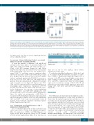
Figure 3. Lower number of tumor-infiltrating T cells is associated with poor survival in primary testicular lymphoma (PTL). (A) Representative images (high, inter- mediate, low) from mIHC analysis of PTL tumor-associate macrophages probed with a 4-plex panel of T-cell markers. Blue: CD3; red: CD8; white: CD4; green: CD56; gray: DAPI. Scale bars 50 μm (upper panel) and 20 mm (lower panel). Images from individual channels are presented in Online Supplementary Figure S4. (B) Boxplots visualizing the expression of CD3+, CD3+CD4+, and CD3+CD8+ lymphocytes in the three groups based on the T-lymphocyte signature (1: poor prognosis, 2: intermedi- ate, and 3: better prognosis). Statistical significance was determined by Kruskall-Wallis test.
ture genes were low (data not shown), suggesting that they are not clinically relevant.
Low amount of tumor-infiltrating T cells is associated with poor outcome in patients with PTL
To study the presence of different T-cell and NK-cell subtypes in the tumor milieu, we performed mIHC with CD3, CD4, CD8, and CD56 cell surface markers on a TMA of 60 PTL patients (Figure 3A and Online Supplementary Figure S4). The relative levels of tumor-infil- trating CD3−CD56+ cells were very low (median 0.3%, range 0.0-52.7%), and there were no significant differ- ences in the amount of CD56+ cells between the good and poor prognosis groups (data not shown). However, mIHC analysis confirmed significantly (P<0.001) lower propor- tions of CD3+, CD3+CD4+, and CD3+CD8+ lymphocytes in patients with a poor prognosis in comparison to patients with a favorable outcome (Figure 3B). Expression of CD3, CD4, and CD8 at the protein level correlated with the cor- responding gene expressions (Spearman P=0.779, P<0.001; P=0.645, P<0.001; and P=0.768, P<0.001, respec- tively). Furthermore, low amounts of tumor-infiltrating CD3+, CD3+CD4+, and CD3+CD8+ lymphocytes were associated with a poor outcome (Online Supplementary Figure S5), which was independent of IPI (Table 3). Consistent with the findings on the gene expression level, an adverse prognostic impact of the low tumor-infiltrating CD4+ and CD8+ T-cell counts was particularly evident in patients treated with the rituximab-containing regimen (Online Supplementary Figure S6).
Loss of membrane-associated HLA class I and II expression is frequent in PTL
Loss or aberrant expression of HLA molecules may cause tumor cells to escape from immunosurveillance. Here, the expression of HLA I and HLA II genes was lower in the poor prognosis group of PTL patients (P<0.05) (Online Supplementary Figure S7A). This was evi-
Table 4. HLA and β2M immunohistochemical staining results. β2M HLA-ABC HLA-DR
Negative
Moderately positive
Highly positive
%%%
31.7 18.3 53.3
55.0 53.3 30.0
13.3 28.3 16.7
dent also in the de novo DLBCL cohort (Online Supplementary Figure S7B).
To study the subcellular localization of HLA class I and II, we analyzed HLA-DR, HLA-ABC and β2M, a compo- nent of HLA I, immunohistochemically (Figure 4A). A minority of the PTL cases showed highly positive mem- brane staining for HLA-DR, HLA-ABC, and β2M (17%, 28%, and 13%, respectively) (Table 4). Interestingly, triple-positive membrane staining of HLA I, HLA II and β2M was associated with a higher number of tumor-infil- trating CD3+, CD3+CD4+, and CD3+CD8+ lymphocytes (Figure 4B).
Discussion
The TME plays an important role in lymphoma patho- genesis and patient prognosis.3 In this study, we aimed to characterize the immunological profiles and their associa- tion with outcome in patients with PTL. We found that a gene signature enriched for T-cell genes was associated with outcome in patients treated with chemotherapy and immunochemotherapy. We could further demonstrate that lower amounts of tumor-infiltrating CD4+ and CD8+ T cells was associated with a poor prognosis in patients with PTL. The impact was particularly evident among patients treated with rituximab-containing immunochemotherapy. The limitation of our study is the lack of a proper validation cohort due to the rare nature of
haematologica | 2019; 104(2)
343
