
Page 140 - 2019_02-Haematologica-web
P. 140
S-K. Leivonen et al.
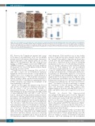
A
B
Figure 4. Loss of membrane-associated HLA class I and II expression is frequent in primary testicular lymphoma (PTL) and correlates with low T-cell infiltration.
(A) Representative images of HLA-ABC, HLA-DR, and β2 microglobulin antibody of PTL tissue sections. The samples were scored as negative (neg), moderately pos- itive (pos), and highly positive according to the membrane staining. Scale bar 50 mm. (B) Boxplots visualizing the correlation of HLA-ABC, HLA-DR and β2M triple- positive membrane staining (either moderate or high membrane staining) with T-cell numbers in PTL (n=14 and n=46 in positive and negative groups, respectively). P-values were determined by Mann-Whitney test.
PTL. However, the T-lymphocyte signature had a prog- nostic impact in an independent cohort of de novo DLBCL patients treated with immunochemotherapy, demonstrat- ing the importance of the signature genes also in other aggressive B-cell lymphomas. Our data extend previous findings on DLBCL patients treated with CHOP and R- CHOP-like regimens.16-18 Together, the results emphasize the important role of the T-cell inflamed TME in regulat- ing therapy resistance in PTL.
T lymphocytes, mostly comprising CD4+ and CD8+ T cells, play a major role in cell-mediated immunity. Lymphoma cells have been shown to escape immunosur- veillance due to loss of expression or mislocalization of HLA I and II molecules.7-11,19 We found that reduced mem- branous staining of HLA I and II molecules and β2M cor- related with lower T-cell infiltration, implying that defects in HLA complexes may impair the recruitment of the tumor-infiltrating T-cell subsets.
Indeed, our data suggest that immune escape does not only provide a mechanism for lymphoma pathogenesis, but also plays a role in promoting resistance to immunochemotherapy. We propose that lymphomas with inflammatory profile characterized by high content of tumor-infiltrating CD4+ and CD8+ T cells, “the hot tumors”, display pre-existing antitumor immune response. In response to therapy, and particularly rituximab-contain- ing regimen, tumor-infiltrating T cells are stimulated fur- ther to participate in immune response against lymphoma cells. In contrast, lymphomas that lack T-cell infiltration, “the cold tumors”, reflect the absence of pre-existing anti- tumor immunity and have a lower likelihood of having an optimal response to therapy. Consistent with our hypoth- esis, it has been shown that many chemotherapeutic drugs, including cyclophosphamide and doxorubicin, which are the main components in the CHOP regimen, can activate anti-tumor immune response by increasing immunogenicity of malignant cells as well as by directly relieving immunosuppressive networks.20 Rituximab and
other therapeutic CD20 antibodies can, in turn, further promote a long-term anti-tumor immune response, called the “vaccinal” effect, which is dependent on the presence of both CD4+ and CD8+ lymphocytes.21-23 Further studies should aim to characterize in more detail the underlying mechanisms for the loss of T-cell trafficking and infiltra- tion. For example, differences in the mutational density between the T-cell inflamed “hot” and non-inflamed “cold” tumors might explain the loss of T cells in a subset of tumors. Additional gene expression profiling studies could provide information as to which genes and molecu- lar pathways are differentially expressed or activated in the T-cell inflamed and non-inflamed tumors, and thus might mediate T-cell exclusion from the TME. For exam- ple, in melanoma and bladder cancer, the Wnt/β-catenin pathway has been shown to be causal in preventing T-cell activation and trafficking into the tumor microenviron- ment.24,25 In addition, it would be interesting to examine the distribution of other cell lineages and their pheno- types, and determine whether PTLs express other immunoregulatory molecules, including PD-L1, LAG-3 or IDO-1 and IDO-2, which can be targeted by novel thera- pies.
Recently, we showed that tumor-associated macrophages (TAMs) play a major role in PTL.26 High infil- tration of PD-L1+CD68+ TAMs was associated with favor- able survival and correlated with CD4+ and CD8+ T cells positive for PD1. In addition, both PD-L1+ TAMs and PD-1+ T cells emerged as independent indicators of sur- vival for the patients with PTL. The interaction of PD-L1+ TAMs and PD-1+ T cells may modify the TME in PTL, or otherwise promote an anti-tumor immune response fol- lowing immunochemotherapy.
The poor outcome associated with a low tumor-infil- trating T-cell content might provide the objective for ther- apeutic interventions. Cancer immunotherapy, especially protocols aiming to activate T-cell-mediated anti-tumor responses, and T-cell trafficking into the non-inflamed
344
haematologica | 2019; 104(2)
