
Page 121 - 2019_02-Haematologica-web
P. 121
ASNase hypersensitivities are IgG/IgE dependent
ABCD
EFGH
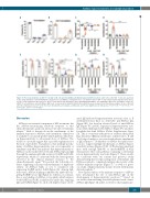
Figure 5. The in vivo depletion of CD4+ T cells but not B cells prevents ASNase-mediated hypersensitivity reactions. (A) B cells or (B) CD4+ T cells were depleted in vivo using anti-CD19 or anti-CD4 mAb three days prior to each ASNase sensitization dose. Less than 5% of B or CD4+ T cells remained after depletion in the blood. (C) The rectal temperature AUC after B or CD4+ T-cell depletion was determined after challenging with ASNase. (D) Anti-ASNase IgG levels, (E) ASNase activity, (F) mMCP-1 concentrations, and anti-ASNase IgE levels, as assessed by (G) ELISA and (H) flow cytometry, were measured from the plasma samples of mice collected after the challenge. A total of 5 or 10 mice were included in each analysis, as indicated, and P value significance is indicated as * for P<0.05, ** for P<0.01, *** for P<1x10⁻³, and **** for P<1x10⁻⁴.
Discussion
ASNase is an essential component of ALL treatment, but the ASNase-neutralizing antibody response to this chemotherapeutic agent can increase the risk of leukemia relapse.13 Little is known about the mechanism of the immune response to ASNase, and currently no clinical test is available to accurately predict which patients will devel- op hypersensitivity to subsequent doses of ASNase. The results of our study suggest that a possible explanation for the lack of predictive biomarkers is that multiple mecha- nisms of ASNase hypersensitivity can occur separately or simultaneously. Our data indicate that hypersensitivity to ASNase can be mediated by both anti-ASNase IgG and IgE through the immunoglobulin receptors FcγRIII and FcεRI, respectively, which is consistent with the heterogeneity observed in the clinical immune response to the chemotherapeutic agent.2 We provide the following evi- dence supporting our proposed mechanisms of hypersen- sitivity: (1) ASNase binds ex vivo to sensitized cells expressing FcγRIII and/or FcεRI (Figure 1B). Among IgE- leukocytes, ASNase binding is inhibited by antibodies tar- geting FcγRIII but not IgE (Figure 2), whereas, in contrast, the ex vivo binding of ASNase to basophils, which express both FcγRIII and FcεRI, is inhibited by antibodies targeting IgE and FcγRIII (Figure 2). (2) B cell-depleted mice devel-
oped IgE-mediated hypersensitivity reactions; that is, B cell-depleted mice have no detectable anti-ASNase IgG (Figure 5D), but develop elevated levels of anti-ASNase IgE (Figure 5G and H), experience hypersensitivity reac- tions when challenged with ASNase (Figure 5C) and have basophils that bind ASNase (Online Supplementary Figure S8). This is consistent with previous evidence that partial Rag deficiency (Omenn syndrome), which leads to B and T-cell oligoclonality, is associated with relative enrichment of IgE responses.29 (3) Markers of ASNase basophil activa- tion also support multiple mechanisms of ASNase hyper- sensitivity in mice (Figure 6C-D). (4) Pretreating sensitized mice with anti-IgE (EM-95) or anti-FcγRIIB/III (2.4G2) demonstrates that each pathway is playing a substantial role in hypersensitivity reactions (Figure 7). These results are consistent with our current and previous study demonstrating that receptor antagonists of histamine and PAF are required to completely mitigate the severity of the ASNase immune response (Figure 7),21 as well as a previ- ous study of hypersensitivity in mice treated with insulin- derived peptides.24
Few clinical studies of the immune response to ASNase have investigated the role of anti-ASNase IgE on the development of hypersensitivity reactions.14-16 Based on our results demonstrating detectable plasma levels of anti- ASNase IgE (Figure 1E and F), we believe that antigen-spe-
haematologica | 2019; 104(2)
325
