
Page 120 - 2019_02-Haematologica-web
P. 120
S. Rathod et al.
ABCD
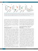
Figure 4. ASNase-sensitized mice develop hypersensitivity reactions when challenged. The rectal temperature of sensitized and non-sensitized mice was monitored for 2 hours after the ASNase challenge on Day 24. (A) Sensitized mice experienced a drop in rectal temperature that was quantified by estimating the AUC of the rectal temperature versus time curve. (B) Sensitized mice had elevated levels of mMCP-1 and (C) low ASNase drug levels compared to non-sensitized mice. (D) ASNase binds to B cells, neutrophils, macrophages/monocytes, basophils, and T cells after challenging with ASNase on Day 24. A total of 5 or 20 mice were included in each analysis, as indicated, and P value significance is indicated as * for P<0.05, ** for P<0.01, *** for P<1x10⁻³, and **** for P<1x10⁻⁴.
cell depletion relative to naïve mice (Online Supplementary Figure S8A-B, P<1x10⁻⁴), but at about half of the frequency relative to sensitized mice with no cell depletion (Figure 4D). No binding of ASNase to B cells or neutrophils was detected after CD4+ T-cell or B-cell depletion (Online Supplementary Figure S8A-B, P>0.05). The data suggests that binding of ASNase to B cells, neutrophils, or macrophages/monocytes may not correlate with the onset of ASNase hypersensitivity, whereas the binding of ASNase to basophils may be a useful marker of ASNase hypersensitivity.
A basophil activation test (BAT) can detect IgG and/or IgE ASNase hypersensitivity
A basophil activation test was used to determine if ASNase-induced activation can be mediated and detected via both ASNase immune complex binding to FcγRIII and free ASNase binding to cell-associated IgE. Blood samples from sensitized and non-sensitized mice were collected on Day 23 of the sensitization protocol (Online Supplementary Figure S3A) and incubated with ASNase, RPMI-medium (negative control), EM-95 (positive control for IgE-mediated activation, Figure 6A) or 2.4G2 (positive control for IgG- mediated activation, Figure 6B). Samples from naïve mice showed no ASNase-mediated basophil activation (Figure 6C-D); however, ASNase-sensitized mice showed an upregulation of CD200R1 and a downregulation of CD200R3 relative to naïve mice (Figure 6C-D, P<0.01), sug- gesting that the activation of basophils and possibly the onset of ASNase hypersensitivities may be mediated by both pathways of anaphylaxis. We anticipated that anti- ASNase IgG may interfere with the binding of ASNase to cell-associated IgE.26 Consistent with that hypothesis, after washing the samples with mouse plasma and removing anti-ASNase IgG antibodies, CD200R3 was no longer downregulated after incubation with ASNase (Figure 6D, P>0.05), whereas a significant upregulation of CD200R1 after removing anti-ASNase IgG was measured relative to sensitized basophils with no antibody removal and to naïve control samples (Figure 6C, P<0.01).
To determine if the BAT can distinguish between sensi- tized and non-sensitized samples from mice with ASNase exposure, we collected blood samples after a single sensiti-
zation dose of ASNase on Day 9 and challenged the mice with IV ASNase on Day 10 (Online Supplementary Figure S3C). We detected no basophil activation by BAT (Online Supplementary Figure S9A-B) or ex vivo ASNase binding to basophils (Online Supplementary Figure S9C). Consistent with these results, mice did not develop hypersensitivity reac- tions upon ASNase challenge (Online Supplementary Figure S9D). Taken together, our data suggest that the binding of ASNase to basophils and ASNase-mediated basophil activa- tion correlate with the onset of ASNase hypersensitivity.
In vivo blocking with EM-95 or 2.4G2 suggests multiple mechanisms of ASNase-induced hypersensitivity
In vivo blocking experiments with EM-95 or 2.4G2 were performed to determine whether ASNase hypersensitivity occurs via both the FcεRI/IgE- and FcγRIII/IgG-mediated pathways as suggested by our BAT data. Mice were pre- treated with either mAb on Day 23 and challenged the next day with ASNase. Mice receiving EM-95 or 2.4G2 were partially protected from hypersensitivity compared to sensitized mice (Figure 7, P<1x10⁻³). The results suggest that ASNase hypersensitivity occurs via IgG- and IgE- mediated mechanisms and that hypersensitivity after pre- treatment with EM-95 was due to PAF release and that after 2.4G2 was due to histamine release.20,24 We, there- fore, tested this hypothesis by pretreating mice with CV- 6209 or triprolidine after EM-95 or 2.4G2 administration, respectively, or with each receptor antagonist alone or in combination with no blocking antibody. Consistent with our hypothesis, mice receiving EM-95 or 2.4G2 as well as the appropriate pretreatment medication were completely protected from ASNase hypersensitivity (Figure 7, P>0.05). Pretreating with CV-6209 or triprolidine alone yielded similar results as either blocking antibody (Figure 7, P>0.05), and ASNase hypersensitivity was completely suppressed in sensitized mice pretreated with CV-6209 and triprolidine (Figure 7, P>0.05). Our results indicate that both pathways of hypersensitivity simultaneously play a role in ASNase-mediated reactions and that recep- tor antagonists of both histamine and PAF are required to completely block the severity of the hypersensitivity reac- tion in our mouse model.
324
haematologica | 2019; 104(2)
