
Page 122 - 2019_02-Haematologica-web
P. 122
S. Rathod et al.
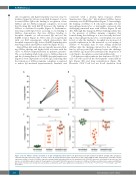
cific recognition and hypersensitivity reactions may be mediated in part by cell-associated IgE. In support of a role for anti-ASNase IgE in the binding or recognition of free ASNase and not ASNase immune complexes, we found that blocking IgE with EM-95 decreases the binding of ASNase to sensitized basophils (Figure 2). Furthermore, removing soluble IgG before assessing ex vivo binding to ASNase demonstrates that free ASNase binding to basophils depends on cell-associated IgE and not the FcγRIII receptor (Figure 2). These data are in agreement with our BAT experiments, which demonstrate that basophils activation is IgE- but not FcγRIII-dependent after removing soluble anti-ASNase antibody (Figure 6C-D).
Anti-ASNase IgG antibodies are typically measured dur- ing ASNase therapy and higher levels associate with the onset of ASNase hypersensitivity in pediatric patients.2 The ex vivo binding of IgE- leukocytes to ASNase (Figure 2) and the downregulation of CD200R3 basophil expression (Figure 6) were dependent on soluble IgG, indicating that the formation of ASNase immune complexes is required to induce IgG-mediated hypersensitivity. Similarly, and
consistent with a murine IgG1 response (Online Supplementary Figure S4),30 IgG-mediated ASNase hyper- sensitivity was FcγRIII-dependent (Figure 7). Interestingly, the binding of ASNase to B cells and basophils, but not macrophages/monocytes or neutrophils, increased after challenging sensitized mice with ASNase (Figure 1B and 4D). Although this change in ASNase binding is likely due to the presence of ASNase immune complexes that formed during the challenge, it is not clear why the bind- ing to macrophages/monocytes or neutrophils was unaf- fected, or why the binding to basophils was increased. A possible explanation for the increased ex vivo binding of ASNase to basophils may be that adding additional ASNase after the challenge allowed for free ASNase to bind to cell-associated IgE, whereas before the challenge anti-ASNase IgG may have neutralized the drug before it could bind to basophil-associated anti-ASNase IgE.
Cell depletion studies indicate that depletion of CD4+ T cells or B cells can block the development of anti-ASNase IgG (Figure 5D) and drug neutralization (Figure 5E). However, CD4+ T-cell but not B-cell depletion (both
AB
CD
Figure 6. Ex vivo ASNase activates basophils in an IgG- and IgE-dependent manner. (A) IgE-mediated basophil activation by EM-95 (anti-IgE mAb) upregulates CD200R1 basophil expression relative to media control. (B) IgG-mediated basophil activation by 2.4G2 (anti-FcγRIIB/III mAB) downregulates CD200R3 basophil expression relative to media. (C) ASNase exposure among sensitized peripheral blood cells upregulates the CD200R1 expression of basophils in the presence and absence of anti-ASNase IgG (no IgG). (D) Downregulation of basophil CD200R3 expression in response to ASNase in ASNase-sensitized mice requires anti-ASNase IgG. A total of 10 mice were included in each analysis, as indicated, and P value significance is indicated as * for P<0.05, ** for P<0.01, *** for P<1x10⁻³, and **** for P<1x10⁻⁴.
326
haematologica | 2019; 104(2)
