
Page 96 - 2019_01-Haematologica-web
P. 96
M. Spagnuolo et al.
AB
CD
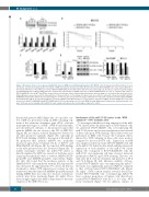
Figure 3. Biological effects of over-expressed miR-17-92 cluster in MYB silenced Philadelphia-positive (Ph+) BV173 cells. (A) (Upper panel) Western blots of a rep- resentative experiment showing specific knockdown of MYB in Doxycycline (Doxy)-treated [24, 48 and 72 hours (h)] BV173-ShMYB cells; (lower panel) qRT-PCR of the indicated members of the miR-17-92 cluster in BV173-ShMYB-Empty Vector (EV) and the miR-17-92 over-expressing cells. Results are expressed as fold changes [mean±Standard Error of Mean (SEM) from three independent experiments] in miRNA expression in BV173-ShMYB-miR-17-92 cells as compared with values in BV173-ShMYB-EV cells. (B) MTT and ATPlite assays; data are the average of three independent experiments, and percentage of cell survival (left panel) and cell via- bility (right panel) were assessed at the indicated times of Doxy treatment. (C) Percentage of S-phase cells over control for untreated or Doxy-treated (48 h) BV173- ShMYB-EV and derivative miR-17-92 over-expressing lines (**P≤0.01). (D) (Left panel) Percentage of Annexin V for untreated or Doxy-treated (96 h) BV173-ShMYB- EV and derivative miR-17-92 over-expressing lines (*P≤0.05). (Middle panel) Western blot of a representative experiment of MYB, uncleaved PARP, BCL-2 and actin protein levels in BV173-ShMYB-EV and BV173-ShMYB-miR-17-92 over-expressing cells, 72 h after MYB silencing. (Right panel) Densitometric analysis by imageJ soft- ware. Actin was used as loading control within the same sample and expressed as fold changes compared to control.
that include putative MBS (Figure 2A). As a positive con- trol, ChIP was performed using an MBS-containing seg- ment of the adenosine deaminase gene (ADA), a known transcriptional target of c-MYB.30 MYB bound efficiently, in both untreated cell lines, to the promoter region that includes MBS#1, the site closest to the TSS of MIR17HG (Figure 2B); in contrast, reduced binding was detected at all other promoter segments (Figure 2B), especially in BV173 cells. Binding of MYB to the region of the miR-17- 92 promoter that includes MBS#1 was markedly decreased upon Doxy treatment (72 h) of BV173- and K562-ShMYB cells (Figure 2B). As expected, MYB binding to the ADA promoter was also decreased (Figure 2B). To further investigate whether the miR-17-92 cluster is directly regulated by MYB we carried out luciferase assay using reporter plasmids with or without MBS#1 (PGL3- prom1353 and ΔMBS#1-prom230, respectively) (Figure 2C, left). We found that the luciferase activity of the ShMYB-BV173 cells transfected with the PGL3-prom1353 was decreased by approximately 33% after a 24 h Doxy treatment to silence MYB expression; in contrast, in cells transfected with the truncated ΔMBS#1-prom230 plasmid lacking MBS#1 there was only a 4% decrease of luciferase activity after Doxy treatment (Figure 2C, right). These data strongly suggest that MYB is important for the tran- scription of the MIR17HG locus.
Involvement of the miR-17-92 cluster in the “MYB addiction” of Ph+ leukemia cells
To investigate whether restoring expression of the miR- 17-92 cluster affects the phenotype of MYB-silenced cells, we generated BV173-ShMYB cells over-expressing the miR-17-92 cluster and assessed proliferation and survival of these cells upon MYB silencing. These studies were not performed in K562 cells because the biological effects induced by MYB silencing in these cells were modest, compared to those in BV173 cells. Expression of MYB was suppressed in Doxy-treated BV173-ShMYB cells and in the miR-17-92 derivative line which exhibited increased expression of each member of the miR-17-92 cluster (Figure 3A). Compared to BV173-ShMYB-EV cells, the miR-17-92 over-expressing cell lines showed increased proliferation (P≤0.01) upon MYB silencing. This was evi- dent after 24 h of Doxy treatment and persisted at 48 h and 72 h (Figure 3B, left). Likewise, viability of Doxy- treated BV173-ShMYB cells over-expressing the miR-17- 92 cluster was significantly increased (P≤0.01) compared to that of Doxy-treated BV173-ShMYB-EV cells (Figure 3B, right). DNA content analysis revealed that Doxy- treated BV173-ShMYB cells over-expressing miR-17-92 have a greater proportion of S-phase cells than Doxy- treated BV173-ShMYB-EV cells (12% vs. 6% after 48 h Doxy treatment) (Figure 3C). In addition, cultures of
86
haematologica | 2019; 104(1)
