
Page 77 - 2019_01-Haematologica-web
P. 77
HbF induction in AML/MDS and outcome prediction
A
B
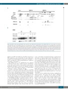
Figure 7. Decitabine induces gene demethylation and expression of the erythroid-specific transcription factor GATA1 in K562 cells. (A) Schematic representation of the GATA1 gene locus. The GATA1 gene is depicted as a black line from 5’ (left) to 3’ (right) with exons represented as boxes. CpG density (CpGs) is indicated by vertical bars. Transcription factor (TF) binding sites were determined through combined TF chromatin immunoprecipitation sequencing (ChIP-seq) experiments from different tissues/cell lines published by the ENCODE consortium. Likewise, activating histone marks H3K9ac and H3K4me3 are taken from ChIPseq experiments in untreated K562 cells which were published and made publicly available from the ENCODE consortium. DNA methylation was assessed by enrichment through methyl- CpG immunoprecipitation sequencing experiments in untreated K562 cells (K562 untr.) and in K562 cells treated with decitabine (K562 DAC). (B) K562 cells were treated with three 24 h pulses of 100 nM or 200 nM decitabine and harvested on days 3 and 6. PMA 5 nM was added to the culture media for the final 48 h. Immunoblotting of whole cell lysates with rat anti-GATA1. Immunoblotting with mouse anti-beta-actin was performed to control for lane loading.
inhibitors in MDS and AML has re-kindled an interest in HbF regulation in vivo. Specifically, we and others have recently described HbF induction in patients with MDS or AML receiving the DNA methyltransferase inhibitors azacitidine or decitabine.14-16,29
Since HMAs have to be administered over repeated treatment cycles in order to induce responses (which then occur only in about half of the patients), early markers predicting subsequent outcome are urgently needed in order to better advise patients whether to continue HMA treatment or switch to alternative therapy.
In order to investigate induction of HbF as a potential predictor of outcome, we chose the time point at the end of two courses of decitabine treatment, i.e. approximately 12 weeks after starting treatment, as the most meaningful: it disclosed the first robust HbF induction (Figure 1A), and was still early enough to be useful for predicting later out- come (whereas information at later time points would be a "self-fulfilling prophecy" regarding response and sur- vival). Indeed, higher HbF at this time point was associat- ed with overall objective responses and, particularly in MDS patients, with improvement of the different hematopoietic lineages after four courses of treatment. Notably, MDS patients with elevated HbF after two
course of decitabine treatment had longer overall survival and trends to longer progression-free and AML-free sur- vival. For AML patients, the median overall survival was also nominally longer in this group, but the difference was not statistically significant (possibly because at that time point, half of the AML patients had already died). Time point optimization in AML patients therefore appears nec- essary, particularly in view of the presently used decitabine treatment schedules of drug administration 5 or 10 days every 4 weeks. It was encouraging to note that higher HbF levels after two courses of treatment were associated with platelet doubling after one course, one of the few established early dynamic predictors of outcome with HMAs.30,31
In order to assess whether these results are applicable in clinical practice, and given the fact that a different decitabine dose is commonly used in MDS and AML patients at present, these results warrant validation in a larger cohort of patients treated with current dosing schemes. For this purpose, serial measurements of HbF have recently been performed in decitabine-treated AML patients randomized within the "inDACtion versus induc- tion" Intergroup AML trial 1301 of the EORTC Leukemia Group (NCT02172872).
haematologica | 2019; 104(1)
67
