
Page 75 - 2019_01-Haematologica-web
P. 75
HbF induction in AML/MDS and outcome prediction
and hemin indicated absence of megakaryocytic differen- tiation (Figure 5C).
Transcriptome profiling by mRNA expression arrays in K562 cells revealed the greatest similarity between hemin- and decitabine-treated cells (Figure 6A,B). Notably, tran- scriptome changes of PMA-treated K562 cells were most extensive and clustered most distantly from those of all other treatments (Figure 6A,B). In HEL cells, variable probes also differed considerably between decitabine and PMA treatment (Online Supplementary Figures S8 and S9). Gene ontology analyses of upregulated transcripts consis- tently identified terms related to erythropoiesis and iron metabolism among the top regulated transcript groups in decitabine-treated cells whereas terms related to megakaryocyte lineage differentiation did not show sig- nificance (data not shown).
Among the transcripts upregulated by decitabine, almost all major globin genes could be identified (Figure 6C). Decitabine-treated K562 showed a >7-fold induction of alpha-1/alpha-2-globin transcription whereas PMA treat- ment did not alter alpha-globin transcription. Gamma-glo- bin transcription was induced by all three treatments, with decitabine exhibiting the most pronounced effect. A strong induction was also noted for zeta-globin transcription upon decitabine and hemin, but not PMA, treatment. Similar patterns were noted for HEL (data not shown).
To prove induction of functionally competent HbF mol- ecules in K562 cells by decitabine treatment, we quantified hemoglobin tetramers by cation-exchange chromatogra- phy. In two independent experiments, a reproducible time- and dose-dependent (albeit modest) induction of HbF tetramers was noted (Figure 6D).
Decitabine-induced globin transcription in K562 cells is associated with GATA1 gene demethylation and upregulation
To determine whether transcriptional upregulation of both the hemoglobin gamma 1 (HBG1) and 2 (HBG2) gene was a consequence of direct DNA methylation changes at the locus control region or at direct upstream regulatory regions of both genes, DNA methylation was assessed globally by methyl-CpG immunoprecipitation, as previ- ously described.24 No enrichment could be demonstrated in untreated or in decitabine-treated K562 cells, indicating absence or only very low levels of DNA methylation at the HBG1 and HBG2 promoter and at the locus control region (Online Supplementary Figure S10). The gamma-glo- bin locus is known to be devoid of a CpG island,25 and was shown to be unmethylated when selected CpG sites were interrogated for their methylation status by using methy- lation-sensitive restriction enzymes.26
The presence of activating H3K4me3 and H3K9ac his- tone marks (from K562 data sets of the ENCODE consor- tium27) and absence of the repressive mark H3K27me3 (data not shown) indicated that transcriptional activity was already present in untreated cells at both loci, which could be demonstrated by the elevated HBG1/HBG2 probe intensity in the mRNA expression array data (Figure 6C).
GATA1 has been identified as a key regulatory factor which binds prominently to the locus control region of the beta-globin locus (Online Supplementary Figure S10). In order to understand how HMA treatment might con- tribute to the transcriptional upregulation of HBG1 and HBG2 without altering pre-existing low levels of local DNA methylation in K562 cells, we assessed DNA methy-
A
B
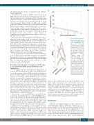
Figure 4. Induced HbF seems to be preferentially derived from non-malignant ery- throid cells. (A) Linear regression model showing the association between HbF induction after four courses of treatment and a decreas- ing percentage of blasts at this time in 17 patients with myelodysplastic syndrome (MDS)/acute myeloid
leukemia (AML). Comparison of HbF levels prior to treatment, after course 2 of decitabine, and at relapse. Three MDS and three AML patients had HbF measurements available at all three time points. In five of the six patients, the initial increase in HbF after course 2 compared to pre-treatment levels became reversed at relapse. In four of these five patients, the level of HbF at relapse was even lower than that at the start of treatment.
(B)
lation at the GATA1 gene locus. Upon decitabine treat- ment, DNA methylation was substantially reduced in K562 cells at the GATA1 promoter region and at an upstream regulatory region (Figure 7A). Western blot analysis of K562 cells after 3 days of decitabine treatment demonstrated a dose-dependent, 2- to 3-fold upregulation of GATA1 protein at day 3 (Figure 7B). After 6 days of treatment, this effect was less pronounced. In contrast, PMA treatment strongly repressed GATA1 expression, an effect only modestly antagonized by decitabine.
Discussion
Clinically meaningful pharmacological induction of developmentally silenced HbF gene expression9,28 is a prime example of what has been termed epigenetic thera- py. However, the broad clinical application of this approach in hemoglobinopathies has been limited by con- cerns of long-term mutagenic effects in these chronic dis- orders. The recent advent of DNA-hypomethylating treat- ment using azanucleoside DNA methyltransferase
haematologica | 2019; 104(1)
65
