
Page 171 - 2019_01-Haematologica-web
P. 171
DOT1L is a therapeutic target in myeloma
8226 cells (Figure 4B). The decreased H3K79me2 levels in these genes in MM cells treated with DOT1L inhibitors were further confirmed by a ChIP-qPCR analysis (Figure 4C). To validate binding of DOT1L to genes marked by H3K79me2, we performed ChIP qPCR and ChIP-seq analysis using an anti-DOT1L antibody. We observed enrichment of DOT1L in the IRF4-MYC signaling genes in both cell lines (Online Supplementary Figure S6).
In addition, qRT-PCR analysis confirmed decreased expression of the 4 genes in MM cell lines treated with DOT1L inhibitors, though our microarray analysis failed
to detect suppression of MYC by SGC0946 in MM.1S cells (Figure 4D). We also confirmed decreased levels of MYC protein in MM cell lines treated with DOT1L inhibitors (Online Supplementary Figure S7), and we found that IRF4 expression was downregulated by DOT1L knockdown in MM cells (Online Supplementary Figure S2A).
DOT1L inhibitors affect immune responses and interferon signaling in MM cells
In addition to their suppressive effects on H3K79me2 and gene expression, DOT1L inhibitors also increased
A
B
C
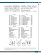
Figure 5, DOT1L inhibition affects immune responses and interferon signaling in MM cells. (A) GO analysis of genes of upregulated (> 1.5-fold) by SGC0946 (1 mM, 6 days) in RPMI-8226 and MM.1S cells. (B) Pathway analysis of genes upregulated by SGC0946 in the indicated MM cell lines. (C) qRT-PCR analysis of interferon- stimulated genes in MM cells treated with the indicated DOT1L inhibitors. Results are normalized to ACTB expression. Shown are means of 3 replications; error bars represent SEMs. *P<0.05; NS: not significant.
haematologica | 2019; 104(1)
161
