
Page 172 - 2019_01-Haematologica-web
P. 172
K. Ishiguro et al.
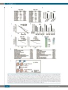
A
B
CD
EF
GH
I
Figure 6. Factors potentially associated with the sensitivity of MM cells to DOT1L inhibitors. (A) Mutations of cancer-related genes detected in KMS-12BM and KMS- 12PE. Genes shared by both cell lines are indicated by gray letters. fsDel: frameshift deletion. (B) qRT-PCR of MYC and IRF4. Results are normalized to ACTB expres- sion. Shown are means of 3 replications; error bars represent SEMs. (C) Results of cell viability assays in KMS-12BM and KMS-12PE cells treated for 2 days with the indicated concentrations of the MYC inhibitor 10058-F4. Results are normalized to cells treated with DMSO. Shown are means of 3 replications; error bars rep- resent SEMs. (D) qRT-PCR analysis of MYC and IRF4 in KMS-12BM and KMS-12PE cells treated with the indicated DOT1L inhibitors (1 mM, 6 days). Shown are means of 3 replications; error bars represent SEMs. (E) Results of cell viability assays in KMS-12PE and U-266 cells subjected to extended DOT1L inhibitor treatment. Cells were treated with SGC0946 (1 mM) or DMSO for 9 days, after which 3x10⁴ cells were placed in a new 6-well plate and cell viabilities were assessed at the indicated times. Results are normalized to cells treated with DMSO. Shown are means of 3 replications; error bars represent SEMs. (F) Heat map showing genes downregulated (> 1.5-fold) by SGC0946 (1 mM, 12 days) in KMS-12PE cells. (G, H) GO (G) and pathway (H) analyses of genes upregulated (> 1.5-fold) by SGC0946 (1 mM, 12 days) in KMS-12PE cells. (I) Hypothesized mechanism underlying the antitumor effect of DOT1L inhibitors in MM cells.
162
haematologica | 2019; 104(1)
