
Page 169 - 2019_01-Haematologica-web
P. 169
DOT1L is a therapeutic target in myeloma
cantly reduced after 6 to 7 days of treatment.17,18 We there- fore performed ChIP-seq analysis of H3K79me2 in RPMI- 8226 and MM.1S cells treated for 3 days with 1 mM SGC0946 or with DMSO, and gene expression microarray analysis with cells treated with the drug for 6 days. ChIP- seq analyses of RPMI-8226 and MM.1S cells respectively identified 4483 and 1590 genes at which H3K79me2 levels were significantly reduced by DOT1L inhibition (Figure 4A, Online Supplementary Tables S1 and S2). Microarray analysis revealed that expression of 912 and 390 genes were downregulated (> 1.5-fold) by SGC0946 in these cells (Online Supplementary Tables S3 and S4). Collectively, we identified 249 genes in RPMI-8226 cells in which both
A
H3K79me2 and expression levels were significantly decreased by SGC0946, while 67 genes were similarly affected in MM.1S cells (Figure 4A). We also identified 13 genes in which both H3K79me2 and expression levels were decreased in both cell lines and 123 genes in which either H3K79me2 levels or their expression levels were decreased in these cell lines (Figure 4A). Among these, we noted that 4 genes associated with IRF4 and MYC signal- ing (MYC, IRF4, PRDM1 and KLF2) were affected in both cell lines (Figure 4A). Visualization of the ChIP-seq data clearly revealed decreased H3K79me2 levels in those 4 genes in both cell lines treated with SGC0946, though dif- ferential peak call analysis failed to detect KLF2 in RPMI-
B
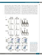
Figure 3. Effects of DOT1L inhibitors on cell cycle and apoptosis in MM cells. (A) Results of cell cycle analysis in MM cells treated with the indicated DOT1L inhibitors (1mM, 6 days). Representative results are shown on the left. Summarized results of 3 replications are shown on the right; error bars represent SEMs. (B) Results of apoptosis assays in MM cell lines treated with the indicated DOT1L inhibitors (1mM, 6 days). The results were confirmed in at least 3 independent experiments, and repre- sentative results are shown (also see Online Supplementary Figure S4).
haematologica | 2019; 104(1)
159
