
Page 168 - 2019_01-Haematologica-web
P. 168
K. Ishiguro et al.
Supplementary Figure S4). Cell cycle and apoptosis were also analyzed at an earlier time point (3 days), and similar results were observed in RPMI-8226 cells (Online Supplementary Figure S5). By contrast, induction of cell cycle arrest and apoptosis were relatively limited in MM.1S cells at this time point, which is consistent with the results of the cell viability assays (Online Supplementary Figure S5, Figure S2A).
DOT1L inhibitors suppress IRF4 and MYC signaling in MM cells
To clarify the molecular mechanism underlying the anti- tumor effects of DOT1L inhibition, we next analyzed H3K79me2 levels and gene expression status in MM cells. Earlier studies showed that 2 to 3 days of DOT1L inhibi- tion resulted in evident depletion of H3K79me2 in tumor cells, while mRNA expression of target genes was signifi-
A
BC
Days
Days
Patient Age Sex Disease Previous treatment
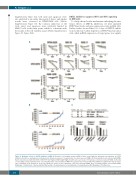
Figure 2. Antitumor effects of DOT1L inhibitors in MM. (A) Results of cell viability assays in MM cell lines treated with the indicated concentrations of DOT1L inhibitors. Results are normalized to untreated cells. Shown are means of 3 replications; error bars represent SEMs. (B) Tumor growth in mice injected with RPMI- 8226 cells pretreated with SGC0946 or EPZ-5676 (left thigh) or DMSO (right thigh). Growth curves are means of 5 replicates; error bars represent SEMs. (C) Results of cell viability assays in primary tumor cells. CD138-positive cells isolated from MM or PCL patients were treated with DOT1L inhibitors (1 μM) for the indicated peri- ods. A summary of the patients is at the top. Shown are means of 3-6 replications; error bars represent SEMs. sPCL: secondary plasma cell leukemia; MPB: Melphalan + Prednisolone + Bortezomib; Bd: Bortezomib + Dexamethasone; Ld: Lenalidomide + Dexamethasone; MP: Melphalan + Prednisolone.
158
haematologica | 2019; 104(1)
